Iron »
PDB 7w1t-7wde »
7wdd »
Iron in PDB 7wdd: Crystal Structure of the P450 BM3 Heme Domain Mutant F87K in Complex with N-Imidazolyl-Hexanoyl-L-Phenylalanine, Styrene and Hydroxylamine
Enzymatic activity of Crystal Structure of the P450 BM3 Heme Domain Mutant F87K in Complex with N-Imidazolyl-Hexanoyl-L-Phenylalanine, Styrene and Hydroxylamine
All present enzymatic activity of Crystal Structure of the P450 BM3 Heme Domain Mutant F87K in Complex with N-Imidazolyl-Hexanoyl-L-Phenylalanine, Styrene and Hydroxylamine:
1.14.14.1; 1.6.2.4;
1.14.14.1; 1.6.2.4;
Protein crystallography data
The structure of Crystal Structure of the P450 BM3 Heme Domain Mutant F87K in Complex with N-Imidazolyl-Hexanoyl-L-Phenylalanine, Styrene and Hydroxylamine, PDB code: 7wdd
was solved by
Y.Jiang,
S.Dong,
Y.Feng,
Z.Cong,
with X-Ray Crystallography technique. A brief refinement statistics is given in the table below:
| Resolution Low / High (Å) | 39.72 / 2.21 |
| Space group | P 1 21 1 |
| Cell size a, b, c (Å), α, β, γ (°) | 58.82, 148.836, 65.336, 90, 100.09, 90 |
| R / Rfree (%) | 19.7 / 22.6 |
Iron Binding Sites:
The binding sites of Iron atom in the Crystal Structure of the P450 BM3 Heme Domain Mutant F87K in Complex with N-Imidazolyl-Hexanoyl-L-Phenylalanine, Styrene and Hydroxylamine
(pdb code 7wdd). This binding sites where shown within
5.0 Angstroms radius around Iron atom.
In total 2 binding sites of Iron where determined in the Crystal Structure of the P450 BM3 Heme Domain Mutant F87K in Complex with N-Imidazolyl-Hexanoyl-L-Phenylalanine, Styrene and Hydroxylamine, PDB code: 7wdd:
Jump to Iron binding site number: 1; 2;
In total 2 binding sites of Iron where determined in the Crystal Structure of the P450 BM3 Heme Domain Mutant F87K in Complex with N-Imidazolyl-Hexanoyl-L-Phenylalanine, Styrene and Hydroxylamine, PDB code: 7wdd:
Jump to Iron binding site number: 1; 2;
Iron binding site 1 out of 2 in 7wdd
Go back to
Iron binding site 1 out
of 2 in the Crystal Structure of the P450 BM3 Heme Domain Mutant F87K in Complex with N-Imidazolyl-Hexanoyl-L-Phenylalanine, Styrene and Hydroxylamine
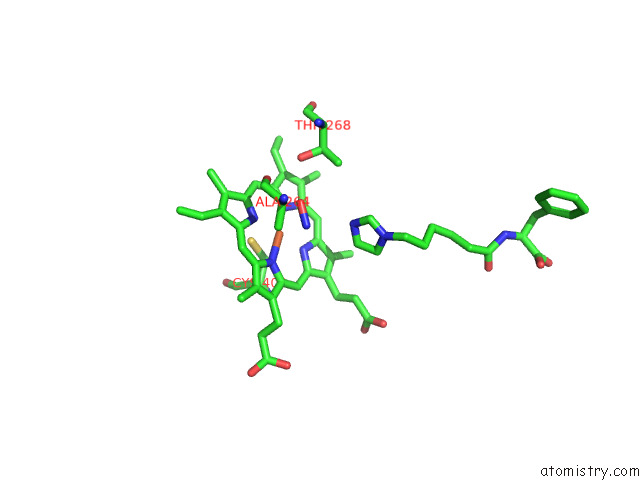
Mono view
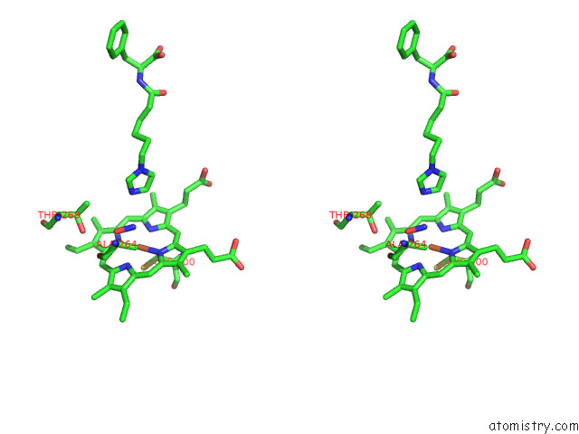
Stereo pair view
Mono view
Stereo pair view
A full contact list of Iron with other atoms in the Fe binding
site number 1 of Crystal Structure of the P450 BM3 Heme Domain Mutant F87K in Complex with N-Imidazolyl-Hexanoyl-L-Phenylalanine, Styrene and Hydroxylamine within 5.0Å range:
|
Iron binding site 2 out of 2 in 7wdd
Go back to
Iron binding site 2 out
of 2 in the Crystal Structure of the P450 BM3 Heme Domain Mutant F87K in Complex with N-Imidazolyl-Hexanoyl-L-Phenylalanine, Styrene and Hydroxylamine
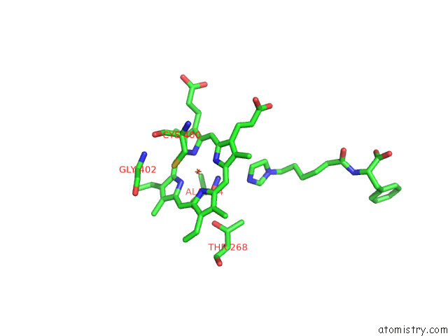
Mono view
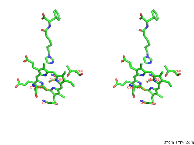
Stereo pair view
Mono view
Stereo pair view
A full contact list of Iron with other atoms in the Fe binding
site number 2 of Crystal Structure of the P450 BM3 Heme Domain Mutant F87K in Complex with N-Imidazolyl-Hexanoyl-L-Phenylalanine, Styrene and Hydroxylamine within 5.0Å range:
|
Reference:
Y.Jiang,
Z.Cong.
Crystal Structure of the P450 BM3 Heme Domain Mutant F87A in Complex with N-Imidazolyl-Hexanoyl-L-Phenylalanine and Hydroxylamine To Be Published.
Page generated: Fri Aug 9 10:05:55 2024
Last articles
Zn in 9IRQZn in 9IYX
Zn in 9J8P
Zn in 9IUU
Zn in 9GBF
Zn in 9G2V
Zn in 9G2L
Zn in 9G2X
Zn in 9G2Z
Zn in 9G2K