Iron »
PDB 7wdg-7xdp »
7x53 »
Iron in PDB 7x53: Cytochrome P450 Monooxygenase
Protein crystallography data
The structure of Cytochrome P450 Monooxygenase, PDB code: 7x53
was solved by
Y.Yan,
C.Zheng,
with X-Ray Crystallography technique. A brief refinement statistics is given in the table below:
| Resolution Low / High (Å) | 23.51 / 3.35 |
| Space group | P 1 21 1 |
| Cell size a, b, c (Å), α, β, γ (°) | 79.831, 55.63, 88.023, 90, 102.94, 90 |
| R / Rfree (%) | 27 / 32.1 |
Iron Binding Sites:
The binding sites of Iron atom in the Cytochrome P450 Monooxygenase
(pdb code 7x53). This binding sites where shown within
5.0 Angstroms radius around Iron atom.
In total 2 binding sites of Iron where determined in the Cytochrome P450 Monooxygenase, PDB code: 7x53:
Jump to Iron binding site number: 1; 2;
In total 2 binding sites of Iron where determined in the Cytochrome P450 Monooxygenase, PDB code: 7x53:
Jump to Iron binding site number: 1; 2;
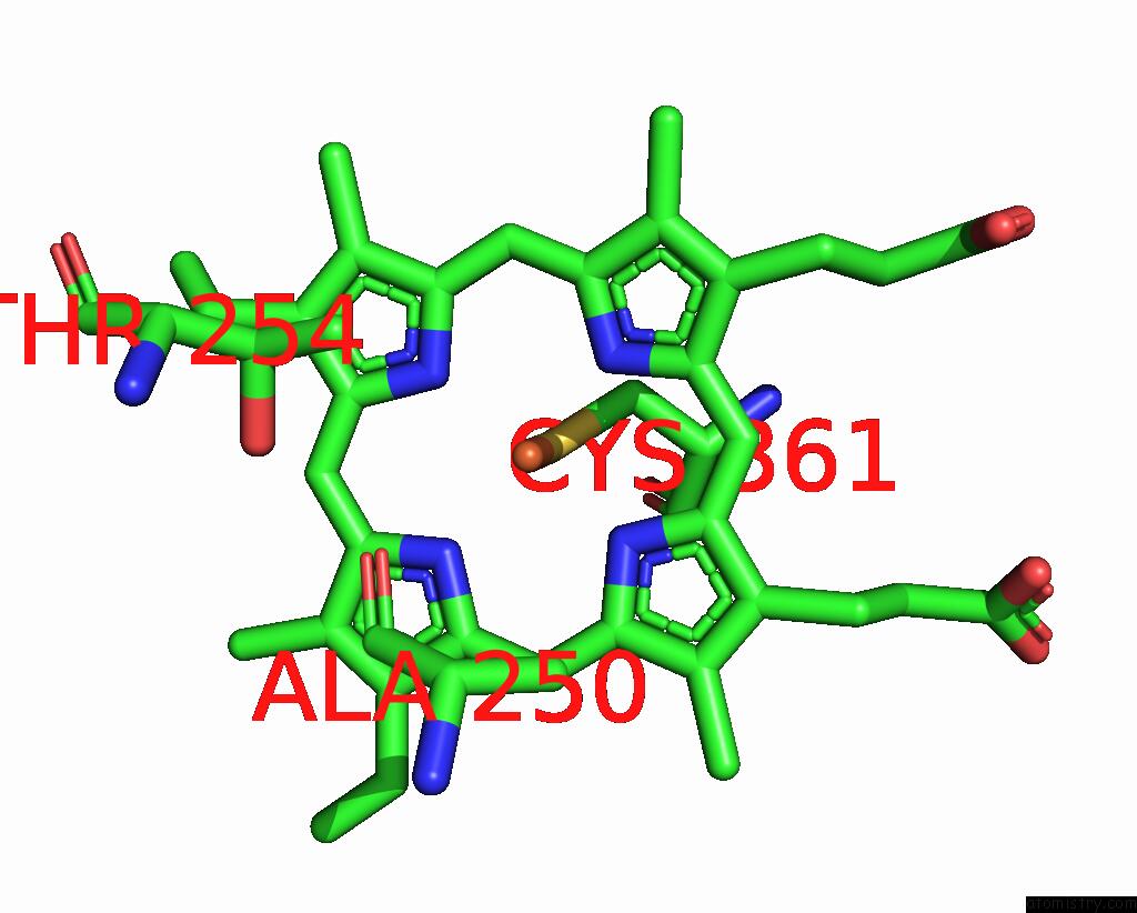
Iron binding site 1 out of 2 in 7x53
Go back to
Iron binding site 1 out
of 2 in the Cytochrome P450 Monooxygenase

Mono view
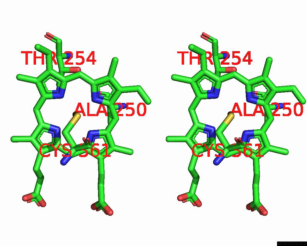
Stereo pair view
Mono view
Stereo pair view
A full contact list of Iron with other atoms in the Fe binding
site number 1 of Cytochrome P450 Monooxygenase within 5.0Å range:
|
Iron binding site 2 out of 2 in 7x53
Go back to
Iron binding site 2 out
of 2 in the Cytochrome P450 Monooxygenase
Mono view
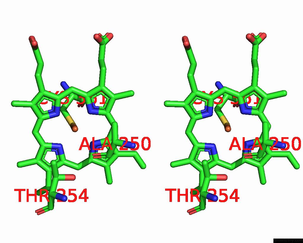
Stereo pair view
Mono view
Stereo pair view
A full contact list of Iron with other atoms in the Fe binding
site number 2 of Cytochrome P450 Monooxygenase within 5.0Å range:
|
Reference:
Y.Yan,
C.Zheng,
S.Wei,
W.Jing,
G.Liang,
G.Cong,
L.Jia,
C.Xiulai,
L.Liming.
Structure of Cytochrome P450 Monooxygenase at 3.35 Angstroms Resolution. To Be Published.
Page generated: Fri Aug 9 10:36:43 2024
Last articles
Cl in 2XZ5Cl in 2XZK
Cl in 2XZC
Cl in 2XYV
Cl in 2XYX
Cl in 2XYR
Cl in 2XZ3
Cl in 2XYW
Cl in 2XYJ
Cl in 2XYQ