Iron »
PDB 7wdg-7xdp »
7xbo »
Iron in PDB 7xbo: Crystal Structure of 10-Dml-Bound Cytochrome P450 Pikc with the Unnatural Amino Acid P-Acetyl-L-Phenylalanine Incorporated at Position 238
Enzymatic activity of Crystal Structure of 10-Dml-Bound Cytochrome P450 Pikc with the Unnatural Amino Acid P-Acetyl-L-Phenylalanine Incorporated at Position 238
All present enzymatic activity of Crystal Structure of 10-Dml-Bound Cytochrome P450 Pikc with the Unnatural Amino Acid P-Acetyl-L-Phenylalanine Incorporated at Position 238:
1.14.15.33;
1.14.15.33;
Protein crystallography data
The structure of Crystal Structure of 10-Dml-Bound Cytochrome P450 Pikc with the Unnatural Amino Acid P-Acetyl-L-Phenylalanine Incorporated at Position 238, PDB code: 7xbo
was solved by
G.B.Li,
Y.J.Pan,
S.Y.Li,
X.Gao,
with X-Ray Crystallography technique. A brief refinement statistics is given in the table below:
| Resolution Low / High (Å) | 32.70 / 2.20 |
| Space group | P 21 21 21 |
| Cell size a, b, c (Å), α, β, γ (°) | 60.119, 108.48, 153.147, 90, 90, 90 |
| R / Rfree (%) | 17.9 / 21.2 |
Iron Binding Sites:
The binding sites of Iron atom in the Crystal Structure of 10-Dml-Bound Cytochrome P450 Pikc with the Unnatural Amino Acid P-Acetyl-L-Phenylalanine Incorporated at Position 238
(pdb code 7xbo). This binding sites where shown within
5.0 Angstroms radius around Iron atom.
In total 2 binding sites of Iron where determined in the Crystal Structure of 10-Dml-Bound Cytochrome P450 Pikc with the Unnatural Amino Acid P-Acetyl-L-Phenylalanine Incorporated at Position 238, PDB code: 7xbo:
Jump to Iron binding site number: 1; 2;
In total 2 binding sites of Iron where determined in the Crystal Structure of 10-Dml-Bound Cytochrome P450 Pikc with the Unnatural Amino Acid P-Acetyl-L-Phenylalanine Incorporated at Position 238, PDB code: 7xbo:
Jump to Iron binding site number: 1; 2;
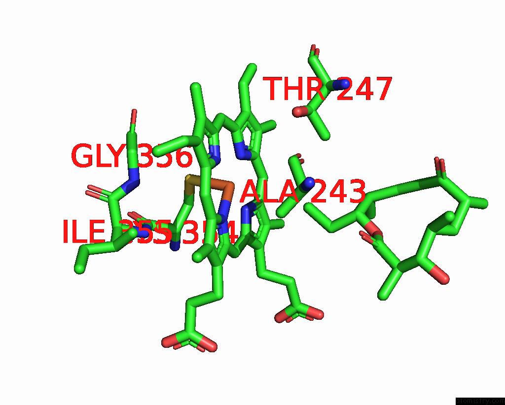
Iron binding site 1 out of 2 in 7xbo
Go back to
Iron binding site 1 out
of 2 in the Crystal Structure of 10-Dml-Bound Cytochrome P450 Pikc with the Unnatural Amino Acid P-Acetyl-L-Phenylalanine Incorporated at Position 238
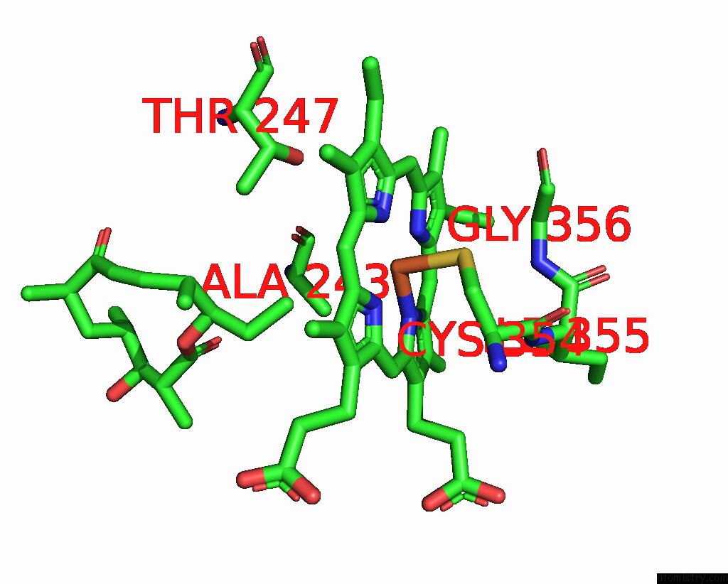
Mono view
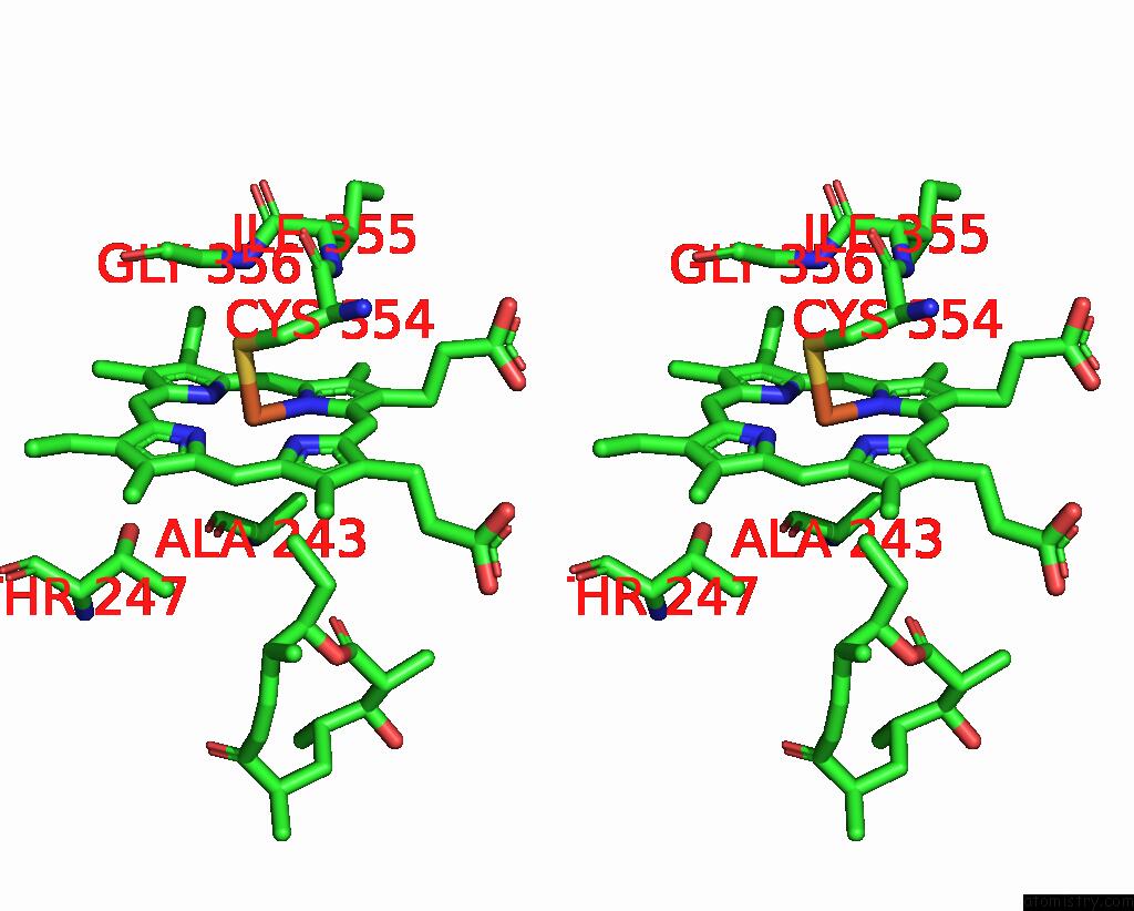
Stereo pair view
Mono view
Stereo pair view
A full contact list of Iron with other atoms in the Fe binding
site number 1 of Crystal Structure of 10-Dml-Bound Cytochrome P450 Pikc with the Unnatural Amino Acid P-Acetyl-L-Phenylalanine Incorporated at Position 238 within 5.0Å range:
|
Iron binding site 2 out of 2 in 7xbo
Go back to
Iron binding site 2 out
of 2 in the Crystal Structure of 10-Dml-Bound Cytochrome P450 Pikc with the Unnatural Amino Acid P-Acetyl-L-Phenylalanine Incorporated at Position 238
Mono view

Stereo pair view
Mono view
Stereo pair view
A full contact list of Iron with other atoms in the Fe binding
site number 2 of Crystal Structure of 10-Dml-Bound Cytochrome P450 Pikc with the Unnatural Amino Acid P-Acetyl-L-Phenylalanine Incorporated at Position 238 within 5.0Å range:
|
Reference:
Y.J.Pan,
G.B.Li,
X.Gao,
S.Y.Li.
New Mechanistic Insight of Cytochrome P450 Pikc Gained From Site-Specific Mutagenesis By Non-Coding Amino Acids Nat Commun 2023.
ISSN: ESSN 2041-1723
Page generated: Fri Aug 9 10:45:47 2024
ISSN: ESSN 2041-1723
Last articles
Zn in 9JYWZn in 9IR4
Zn in 9IR3
Zn in 9GMX
Zn in 9GMW
Zn in 9JEJ
Zn in 9ERF
Zn in 9ERE
Zn in 9EGV
Zn in 9EGW