Iron »
PDB 7xk3-7y9k »
7y5f »
Iron in PDB 7y5f: Crystal Structure of Cmnc in Complex with L-Homoarginine
Protein crystallography data
The structure of Crystal Structure of Cmnc in Complex with L-Homoarginine, PDB code: 7y5f
was solved by
Y.H.Hsiao,
S.J.Huang,
E.C.Lin,
Y.C.Lee,
C.Y.Chang,
with X-Ray Crystallography technique. A brief refinement statistics is given in the table below:
| Resolution Low / High (Å) | 27.89 / 1.52 |
| Space group | I 2 2 2 |
| Cell size a, b, c (Å), α, β, γ (°) | 93.344, 127.514, 139.034, 90, 90, 90 |
| R / Rfree (%) | 18 / 20.4 |
Iron Binding Sites:
The binding sites of Iron atom in the Crystal Structure of Cmnc in Complex with L-Homoarginine
(pdb code 7y5f). This binding sites where shown within
5.0 Angstroms radius around Iron atom.
In total 2 binding sites of Iron where determined in the Crystal Structure of Cmnc in Complex with L-Homoarginine, PDB code: 7y5f:
Jump to Iron binding site number: 1; 2;
In total 2 binding sites of Iron where determined in the Crystal Structure of Cmnc in Complex with L-Homoarginine, PDB code: 7y5f:
Jump to Iron binding site number: 1; 2;
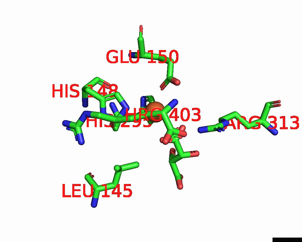
Iron binding site 1 out of 2 in 7y5f
Go back to
Iron binding site 1 out
of 2 in the Crystal Structure of Cmnc in Complex with L-Homoarginine

Mono view
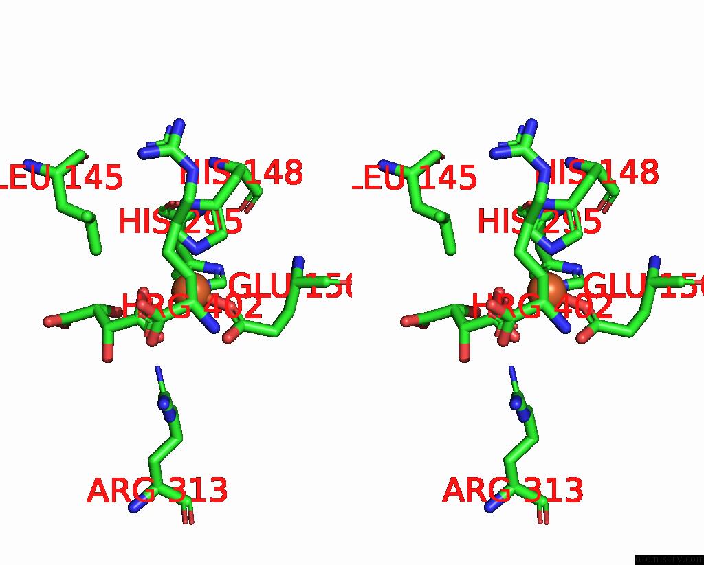
Stereo pair view
Mono view
Stereo pair view
A full contact list of Iron with other atoms in the Fe binding
site number 1 of Crystal Structure of Cmnc in Complex with L-Homoarginine within 5.0Å range:
|
Iron binding site 2 out of 2 in 7y5f
Go back to
Iron binding site 2 out
of 2 in the Crystal Structure of Cmnc in Complex with L-Homoarginine
Mono view
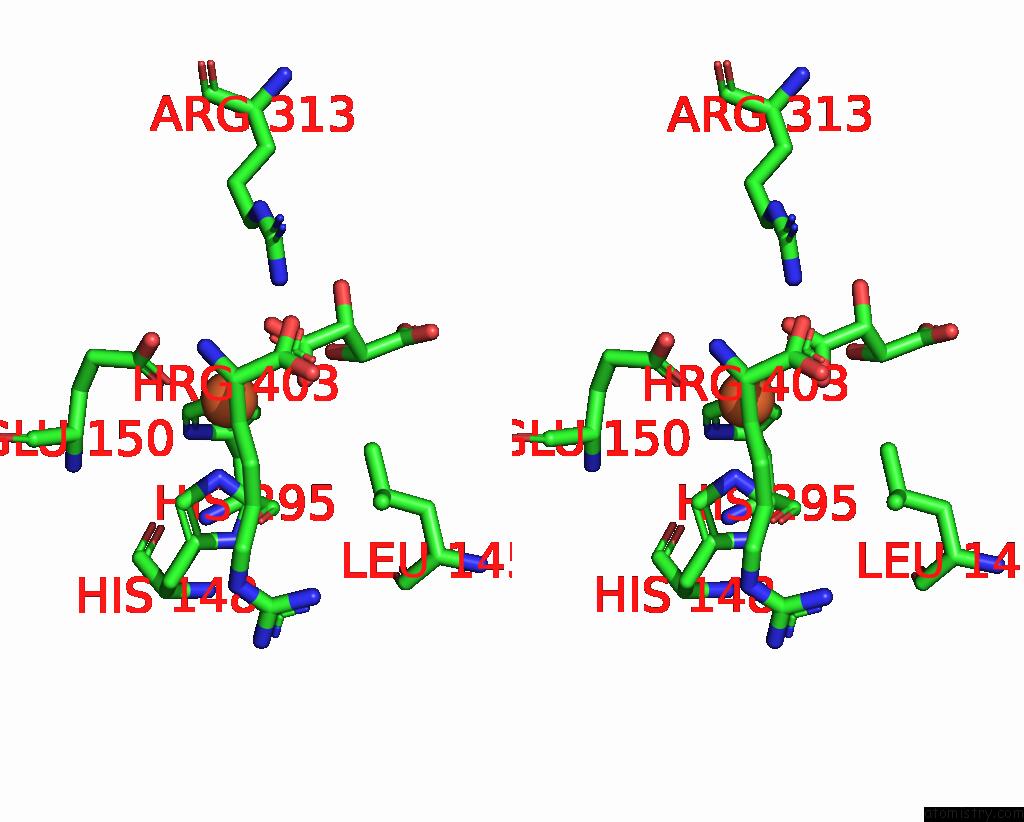
Stereo pair view
Mono view
Stereo pair view
A full contact list of Iron with other atoms in the Fe binding
site number 2 of Crystal Structure of Cmnc in Complex with L-Homoarginine within 5.0Å range:
|
Reference:
Y.H.Hsiao,
S.J.Huang,
E.C.Lin,
Y.C.Lee,
C.Y.Chang.
Crystal Structure of Cmnc in Complex with L-Homoarginine To Be Published.
Page generated: Thu Aug 7 10:14:31 2025
Last articles
Mg in 1Q6TMg in 1Q6Z
Mg in 1Q78
Mg in 1Q6S
Mg in 1Q6R
Mg in 1Q6Q
Mg in 1Q6O
Mg in 1Q6N
Mg in 1Q3U
Mg in 1Q6L