Iron »
PDB 7y9k-7yzq »
7yvz »
Iron in PDB 7yvz: Structure of Caenorhabditis Elegans Cisd-1/Mitoneet
Protein crystallography data
The structure of Structure of Caenorhabditis Elegans Cisd-1/Mitoneet, PDB code: 7yvz
was solved by
K.Hasegawa,
E.Hagiuda,
A.T.Taguchi,
W.Geldenhuys,
T.Iwasaki,
T.Kumasaka,
with X-Ray Crystallography technique. A brief refinement statistics is given in the table below:
| Resolution Low / High (Å) | 34.35 / 1.70 |
| Space group | P 41 21 2 |
| Cell size a, b, c (Å), α, β, γ (°) | 37.798, 37.798, 82.26, 90, 90, 90 |
| R / Rfree (%) | 16.8 / 18.9 |
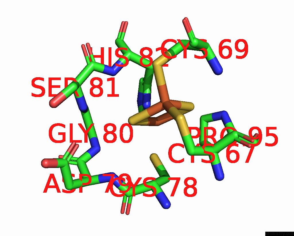
Iron Binding Sites:
The binding sites of Iron atom in the Structure of Caenorhabditis Elegans Cisd-1/Mitoneet
(pdb code 7yvz). This binding sites where shown within
5.0 Angstroms radius around Iron atom.
In total 2 binding sites of Iron where determined in the Structure of Caenorhabditis Elegans Cisd-1/Mitoneet, PDB code: 7yvz:
Jump to Iron binding site number: 1; 2;
In total 2 binding sites of Iron where determined in the Structure of Caenorhabditis Elegans Cisd-1/Mitoneet, PDB code: 7yvz:
Jump to Iron binding site number: 1; 2;
Iron binding site 1 out of 2 in 7yvz
Go back to
Iron binding site 1 out
of 2 in the Structure of Caenorhabditis Elegans Cisd-1/Mitoneet

Mono view

Stereo pair view
Mono view
Stereo pair view
A full contact list of Iron with other atoms in the Fe binding
site number 1 of Structure of Caenorhabditis Elegans Cisd-1/Mitoneet within 5.0Å range:
|
Iron binding site 2 out of 2 in 7yvz
Go back to
Iron binding site 2 out
of 2 in the Structure of Caenorhabditis Elegans Cisd-1/Mitoneet
Mono view

Stereo pair view
Mono view
Stereo pair view
A full contact list of Iron with other atoms in the Fe binding
site number 2 of Structure of Caenorhabditis Elegans Cisd-1/Mitoneet within 5.0Å range:
|
Reference:
J.R.Boos,
H.N.Jandrain,
E.Hagiuda,
A.T.Taguchi,
K.Hasegawa,
B.L.Fedun,
S.J.Taylor,
S.M.Elad,
S.E.Faber,
T.Kumasaka,
T.Iwasaki,
W.J.Geldenhuys.
Structure and Biological Evaluation of Caenorhabditis Elegans Cisd-1/Mitoneet, A Klp-17 Tail Domain Homologue, Supports Attenuation of Paraquat-Induced Oxidative Stress Through A P38 Mapk-Mediated Antioxidant Defense Response Advances in Redox Research V. 6 00048 2022.
DOI: 10.1016/J.ARRES.2022.100048
Page generated: Fri Aug 9 12:28:59 2024
DOI: 10.1016/J.ARRES.2022.100048
Last articles
Zn in 9J0NZn in 9J0O
Zn in 9J0P
Zn in 9FJX
Zn in 9EKB
Zn in 9C0F
Zn in 9CAH
Zn in 9CH0
Zn in 9CH3
Zn in 9CH1