Iron »
PDB 7yzt-7zih »
7z0s »
Iron in PDB 7z0s: Structure of the Escherichia Coli Formate Hydrogenlyase Complex (Anaerobic Preparation, Without Formate Dehydrogenase H)
Other elements in 7z0s:
The structure of Structure of the Escherichia Coli Formate Hydrogenlyase Complex (Anaerobic Preparation, Without Formate Dehydrogenase H) also contains other interesting chemical elements:
| Nickel | (Ni) | 1 atom |
Iron Binding Sites:
Pages:
>>> Page 1 <<< Page 2, Binding sites: 11 - 20; Page 3, Binding sites: 21 - 30;Binding sites:
The binding sites of Iron atom in the Structure of the Escherichia Coli Formate Hydrogenlyase Complex (Anaerobic Preparation, Without Formate Dehydrogenase H) (pdb code 7z0s). This binding sites where shown within 5.0 Angstroms radius around Iron atom.In total 30 binding sites of Iron where determined in the Structure of the Escherichia Coli Formate Hydrogenlyase Complex (Anaerobic Preparation, Without Formate Dehydrogenase H), PDB code: 7z0s:
Jump to Iron binding site number: 1; 2; 3; 4; 5; 6; 7; 8; 9; 10;
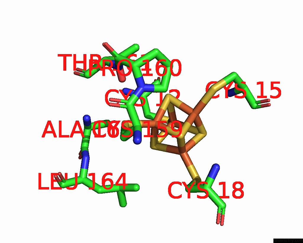
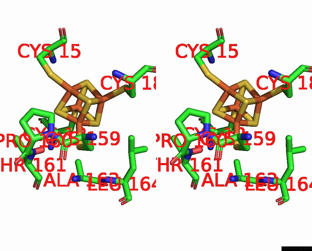
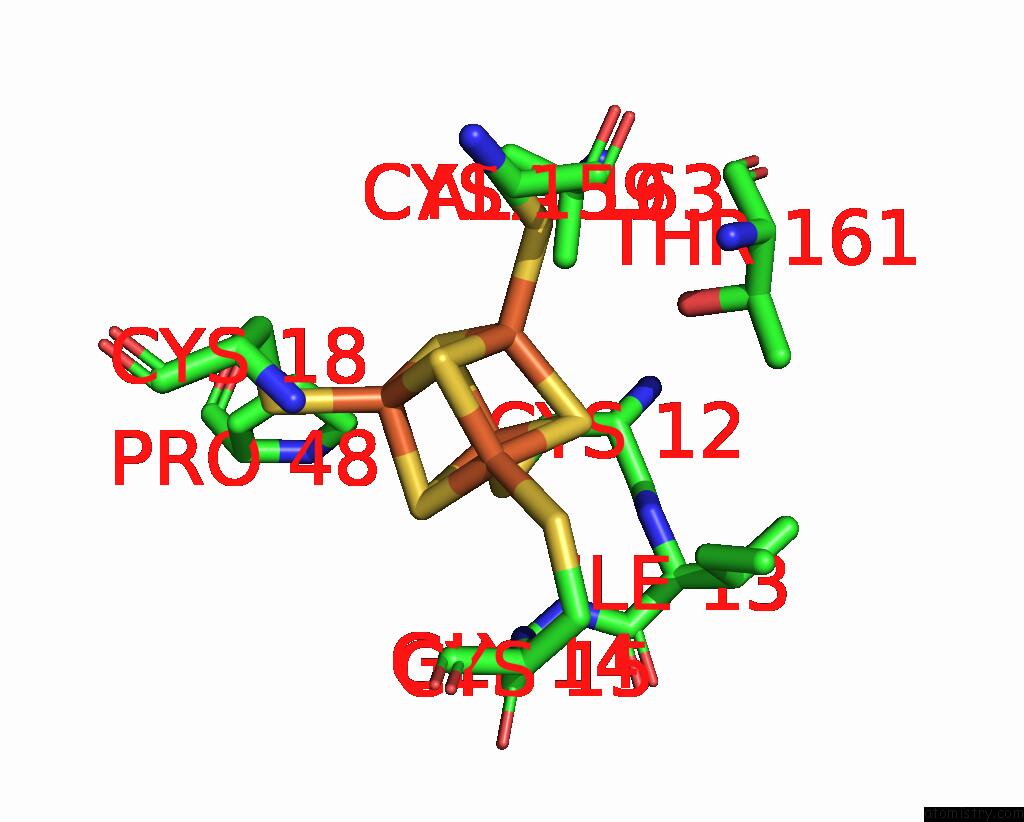
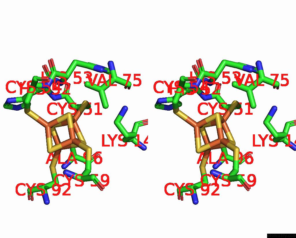
Iron binding site 1 out of 30 in 7z0s
Go back to
Iron binding site 1 out
of 30 in the Structure of the Escherichia Coli Formate Hydrogenlyase Complex (Anaerobic Preparation, Without Formate Dehydrogenase H)
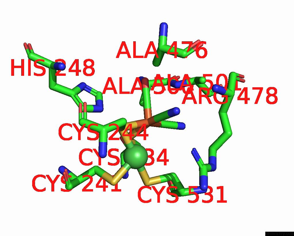
Mono view
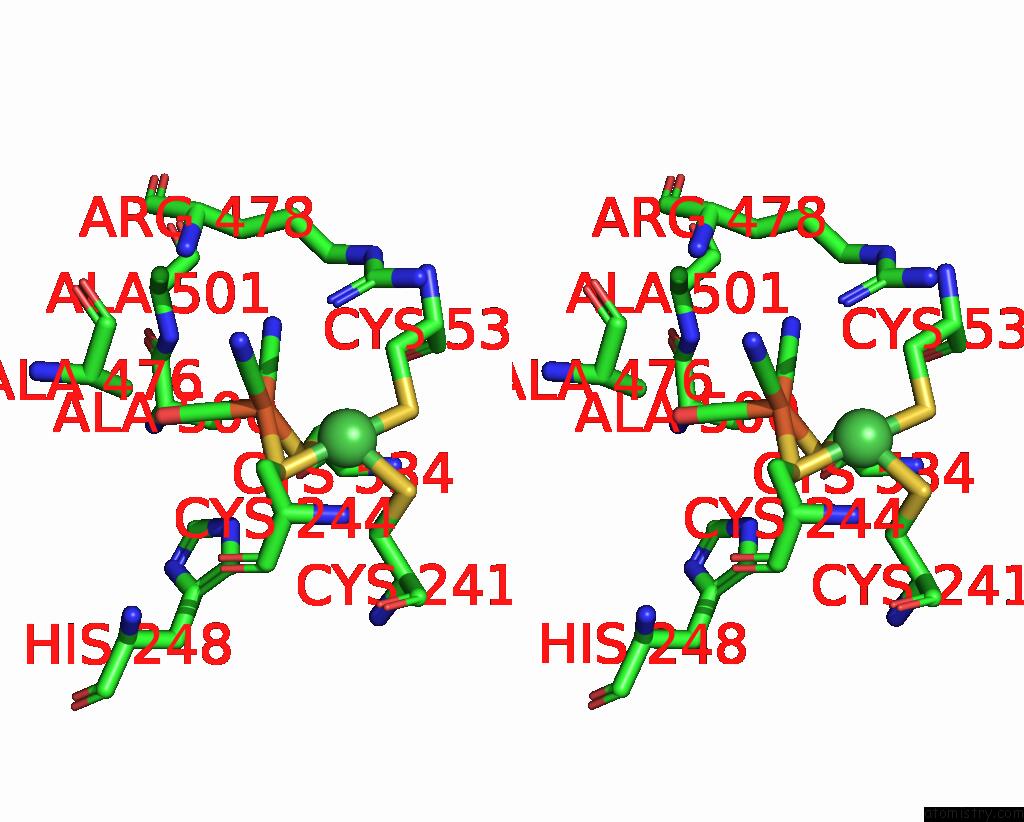
Stereo pair view
Mono view
Stereo pair view
A full contact list of Iron with other atoms in the Fe binding
site number 1 of Structure of the Escherichia Coli Formate Hydrogenlyase Complex (Anaerobic Preparation, Without Formate Dehydrogenase H) within 5.0Å range:
|
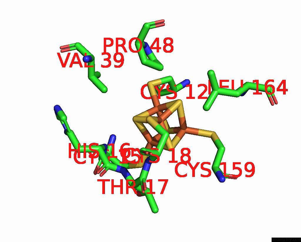
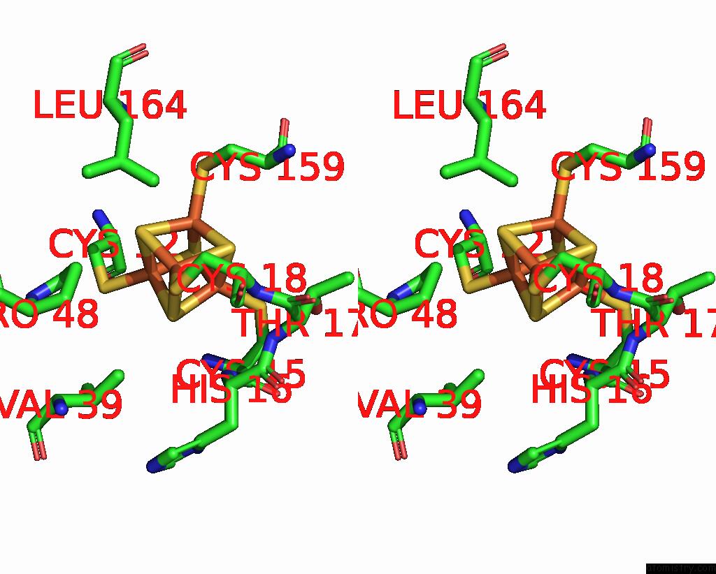
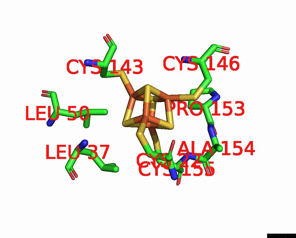
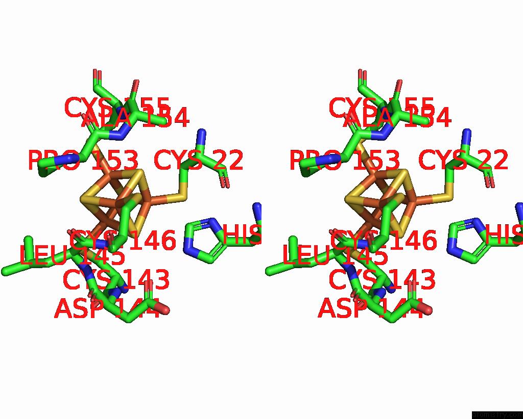
Iron binding site 2 out of 30 in 7z0s
Go back to
Iron binding site 2 out
of 30 in the Structure of the Escherichia Coli Formate Hydrogenlyase Complex (Anaerobic Preparation, Without Formate Dehydrogenase H)
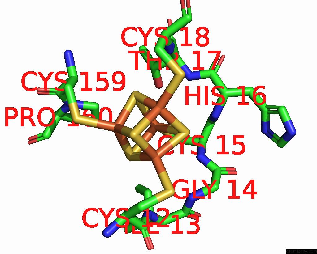
Mono view
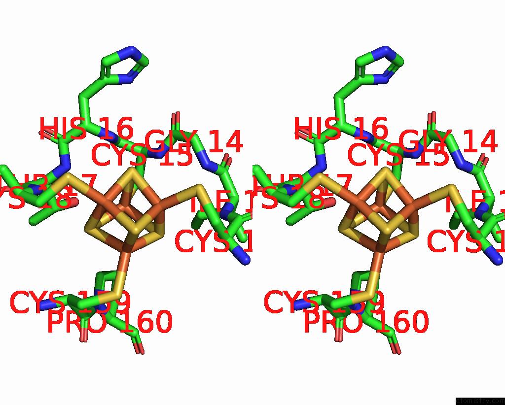
Stereo pair view
Mono view
Stereo pair view
A full contact list of Iron with other atoms in the Fe binding
site number 2 of Structure of the Escherichia Coli Formate Hydrogenlyase Complex (Anaerobic Preparation, Without Formate Dehydrogenase H) within 5.0Å range:
|
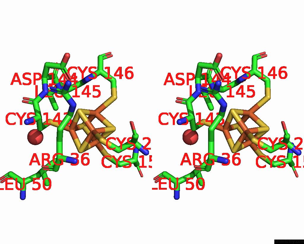
Iron binding site 3 out of 30 in 7z0s
Go back to
Iron binding site 3 out
of 30 in the Structure of the Escherichia Coli Formate Hydrogenlyase Complex (Anaerobic Preparation, Without Formate Dehydrogenase H)
Mono view
Stereo pair view
Mono view
Stereo pair view
A full contact list of Iron with other atoms in the Fe binding
site number 3 of Structure of the Escherichia Coli Formate Hydrogenlyase Complex (Anaerobic Preparation, Without Formate Dehydrogenase H) within 5.0Å range:
|
Iron binding site 4 out of 30 in 7z0s
Go back to
Iron binding site 4 out
of 30 in the Structure of the Escherichia Coli Formate Hydrogenlyase Complex (Anaerobic Preparation, Without Formate Dehydrogenase H)
Mono view
Stereo pair view
Mono view
Stereo pair view
A full contact list of Iron with other atoms in the Fe binding
site number 4 of Structure of the Escherichia Coli Formate Hydrogenlyase Complex (Anaerobic Preparation, Without Formate Dehydrogenase H) within 5.0Å range:
|
Iron binding site 5 out of 30 in 7z0s
Go back to
Iron binding site 5 out
of 30 in the Structure of the Escherichia Coli Formate Hydrogenlyase Complex (Anaerobic Preparation, Without Formate Dehydrogenase H)
Mono view

Stereo pair view
Mono view
Stereo pair view
A full contact list of Iron with other atoms in the Fe binding
site number 5 of Structure of the Escherichia Coli Formate Hydrogenlyase Complex (Anaerobic Preparation, Without Formate Dehydrogenase H) within 5.0Å range:
|
Iron binding site 6 out of 30 in 7z0s
Go back to
Iron binding site 6 out
of 30 in the Structure of the Escherichia Coli Formate Hydrogenlyase Complex (Anaerobic Preparation, Without Formate Dehydrogenase H)
Mono view

Stereo pair view
Mono view
Stereo pair view
A full contact list of Iron with other atoms in the Fe binding
site number 6 of Structure of the Escherichia Coli Formate Hydrogenlyase Complex (Anaerobic Preparation, Without Formate Dehydrogenase H) within 5.0Å range:
|
Iron binding site 7 out of 30 in 7z0s
Go back to
Iron binding site 7 out
of 30 in the Structure of the Escherichia Coli Formate Hydrogenlyase Complex (Anaerobic Preparation, Without Formate Dehydrogenase H)

Mono view
Stereo pair view
Mono view
Stereo pair view
A full contact list of Iron with other atoms in the Fe binding
site number 7 of Structure of the Escherichia Coli Formate Hydrogenlyase Complex (Anaerobic Preparation, Without Formate Dehydrogenase H) within 5.0Å range:
|
Iron binding site 8 out of 30 in 7z0s
Go back to
Iron binding site 8 out
of 30 in the Structure of the Escherichia Coli Formate Hydrogenlyase Complex (Anaerobic Preparation, Without Formate Dehydrogenase H)

Mono view

Stereo pair view
Mono view
Stereo pair view
A full contact list of Iron with other atoms in the Fe binding
site number 8 of Structure of the Escherichia Coli Formate Hydrogenlyase Complex (Anaerobic Preparation, Without Formate Dehydrogenase H) within 5.0Å range:
|
Iron binding site 9 out of 30 in 7z0s
Go back to
Iron binding site 9 out
of 30 in the Structure of the Escherichia Coli Formate Hydrogenlyase Complex (Anaerobic Preparation, Without Formate Dehydrogenase H)

Mono view
Stereo pair view
Mono view
Stereo pair view
A full contact list of Iron with other atoms in the Fe binding
site number 9 of Structure of the Escherichia Coli Formate Hydrogenlyase Complex (Anaerobic Preparation, Without Formate Dehydrogenase H) within 5.0Å range:
|
Iron binding site 10 out of 30 in 7z0s
Go back to
Iron binding site 10 out
of 30 in the Structure of the Escherichia Coli Formate Hydrogenlyase Complex (Anaerobic Preparation, Without Formate Dehydrogenase H)

Mono view
Stereo pair view
Mono view
Stereo pair view
A full contact list of Iron with other atoms in the Fe binding
site number 10 of Structure of the Escherichia Coli Formate Hydrogenlyase Complex (Anaerobic Preparation, Without Formate Dehydrogenase H) within 5.0Å range:
|
Reference:
R.Steinhilper,
G.Hoff,
J.Heider,
B.J.Murphy.
Structure of the Membrane-Bound Formate Hydrogenlyase Complex From Escherichia Coli. Nat Commun V. 13 5395 2022.
ISSN: ESSN 2041-1723
PubMed: 36104349
DOI: 10.1038/S41467-022-32831-X
Page generated: Fri Aug 9 13:00:56 2024
ISSN: ESSN 2041-1723
PubMed: 36104349
DOI: 10.1038/S41467-022-32831-X
Last articles
Zn in 9MJ5Zn in 9HNW
Zn in 9G0L
Zn in 9FNE
Zn in 9DZN
Zn in 9E0I
Zn in 9D32
Zn in 9DAK
Zn in 8ZXC
Zn in 8ZUF