Iron »
PDB 7zxc-8abh »
8a5f »
Iron in PDB 8a5f: Crystal Structure of Deinococcus Radiodurans Endonuclease III-1 R61Q Variant
Protein crystallography data
The structure of Crystal Structure of Deinococcus Radiodurans Endonuclease III-1 R61Q Variant, PDB code: 8a5f
was solved by
P.T.Borges,
F.Rollo,
E.Moe,
with X-Ray Crystallography technique. A brief refinement statistics is given in the table below:
| Resolution Low / High (Å) | 44.82 / 1.38 |
| Space group | C 1 2 1 |
| Cell size a, b, c (Å), α, β, γ (°) | 179.318, 38.04, 36.353, 90, 90.96, 90 |
| R / Rfree (%) | 15.1 / 18 |
Other elements in 8a5f:
The structure of Crystal Structure of Deinococcus Radiodurans Endonuclease III-1 R61Q Variant also contains other interesting chemical elements:
| Magnesium | (Mg) | 2 atoms |
Iron Binding Sites:
The binding sites of Iron atom in the Crystal Structure of Deinococcus Radiodurans Endonuclease III-1 R61Q Variant
(pdb code 8a5f). This binding sites where shown within
5.0 Angstroms radius around Iron atom.
In total 4 binding sites of Iron where determined in the Crystal Structure of Deinococcus Radiodurans Endonuclease III-1 R61Q Variant, PDB code: 8a5f:
Jump to Iron binding site number: 1; 2; 3; 4;
In total 4 binding sites of Iron where determined in the Crystal Structure of Deinococcus Radiodurans Endonuclease III-1 R61Q Variant, PDB code: 8a5f:
Jump to Iron binding site number: 1; 2; 3; 4;
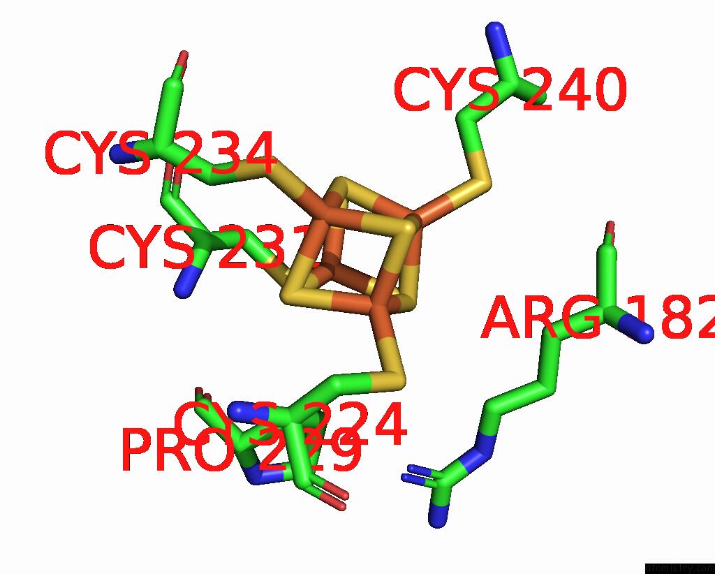
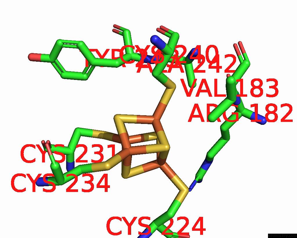
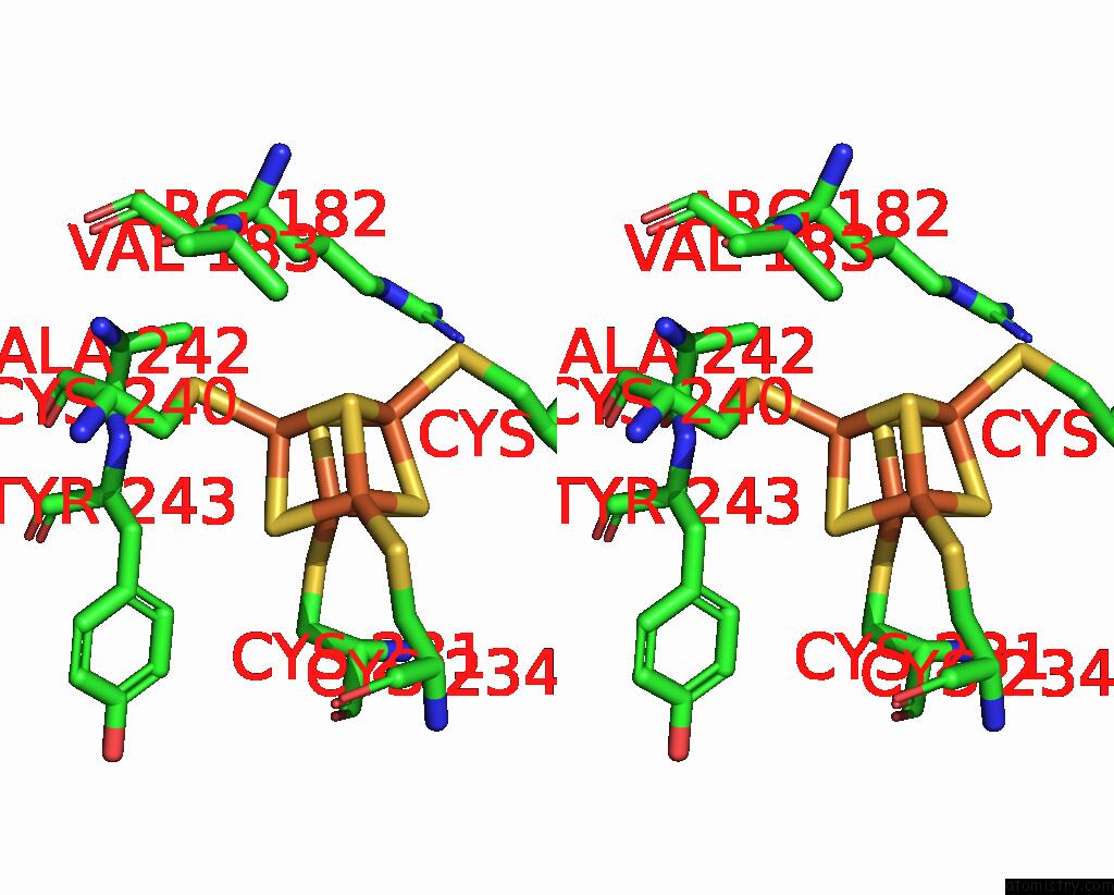
Iron binding site 1 out of 4 in 8a5f
Go back to
Iron binding site 1 out
of 4 in the Crystal Structure of Deinococcus Radiodurans Endonuclease III-1 R61Q Variant
Mono view
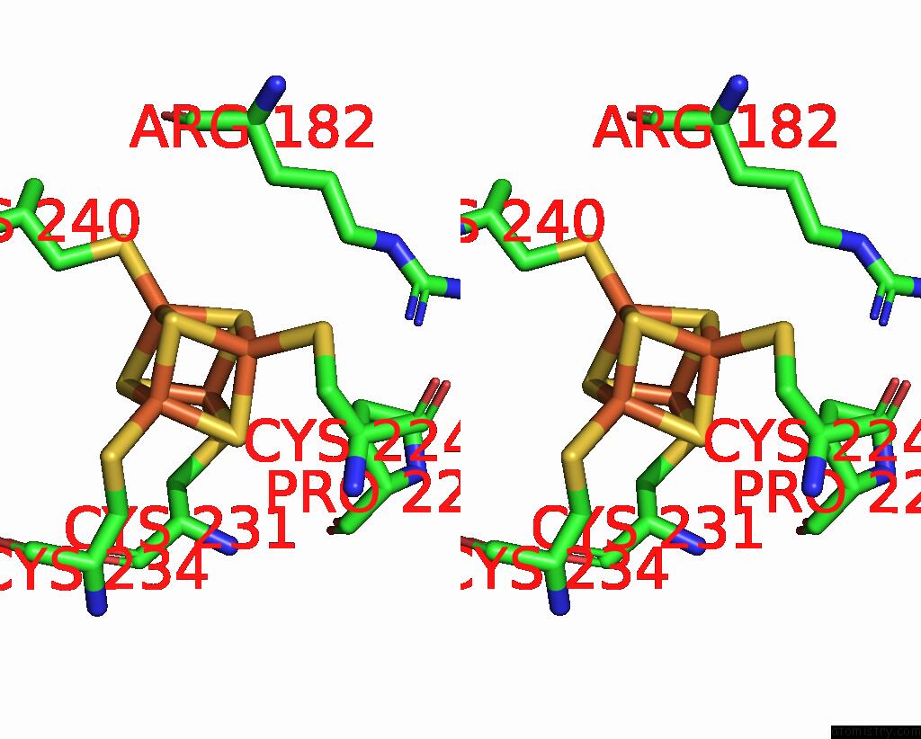
Stereo pair view
Mono view
Stereo pair view
A full contact list of Iron with other atoms in the Fe binding
site number 1 of Crystal Structure of Deinococcus Radiodurans Endonuclease III-1 R61Q Variant within 5.0Å range:
|
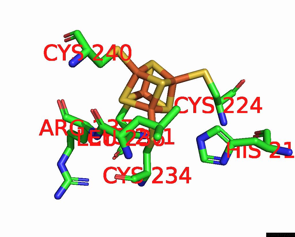
Iron binding site 2 out of 4 in 8a5f
Go back to
Iron binding site 2 out
of 4 in the Crystal Structure of Deinococcus Radiodurans Endonuclease III-1 R61Q Variant
Mono view

Stereo pair view
Mono view
Stereo pair view
A full contact list of Iron with other atoms in the Fe binding
site number 2 of Crystal Structure of Deinococcus Radiodurans Endonuclease III-1 R61Q Variant within 5.0Å range:
|
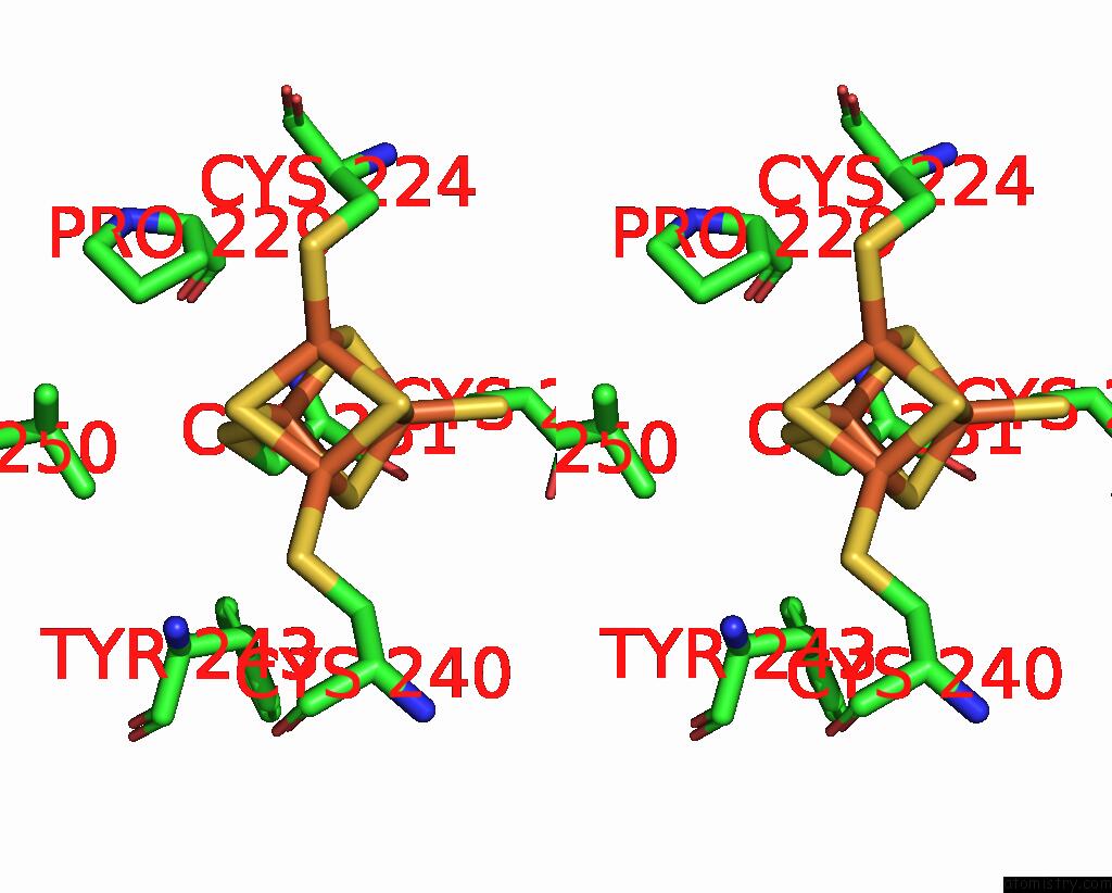
Iron binding site 3 out of 4 in 8a5f
Go back to
Iron binding site 3 out
of 4 in the Crystal Structure of Deinococcus Radiodurans Endonuclease III-1 R61Q Variant

Mono view
Stereo pair view
Mono view
Stereo pair view
A full contact list of Iron with other atoms in the Fe binding
site number 3 of Crystal Structure of Deinococcus Radiodurans Endonuclease III-1 R61Q Variant within 5.0Å range:
|
Iron binding site 4 out of 4 in 8a5f
Go back to
Iron binding site 4 out
of 4 in the Crystal Structure of Deinococcus Radiodurans Endonuclease III-1 R61Q Variant
Mono view
Stereo pair view
Mono view
Stereo pair view
A full contact list of Iron with other atoms in the Fe binding
site number 4 of Crystal Structure of Deinococcus Radiodurans Endonuclease III-1 R61Q Variant within 5.0Å range:
|
Reference:
F.Rollo,
P.T.Borges,
C.M.Silveira,
M.T.G.Rosa,
S.Todorovic,
E.Moe.
Disentangling Unusual Catalytic Properties and the Role of the [4FE-4S] Cluster of Three Endonuclease III From the Extremophile D. Radiodurans. Molecules V. 27 2022.
ISSN: ESSN 1420-3049
PubMed: 35807515
DOI: 10.3390/MOLECULES27134270
Page generated: Thu Aug 7 12:29:59 2025
ISSN: ESSN 1420-3049
PubMed: 35807515
DOI: 10.3390/MOLECULES27134270
Last articles
Mg in 2OQYMg in 2OUP
Mg in 2OUN
Mg in 2OU7
Mg in 2OTG
Mg in 2OSB
Mg in 2OS8
Mg in 2ORI
Mg in 2ORW
Mg in 2ONP