Atomistry » Iron » PDB 7zx5-8abg » 8a90
Atomistry »
  Iron »
    PDB 7zx5-8abg »
      8a90 »

Iron in PDB 8a90: Crystal Structure of Frsh

Protein crystallography data

The structure of Crystal Structure of Frsh, PDB code: 8a90 was solved by N.Schneberger, D.A.Wirtz, M.Cruesemann, G.Hagelueken, with X-Ray Crystallography technique. A brief refinement statistics is given in the table below:

Resolution Low / High (Å) 48.02 / 2.57
Space group P 41 21 2
Cell size a, b, c (Å), α, β, γ (°) 117.715, 117.715, 234.261, 90, 90, 90
R / Rfree (%) 21.7 / 26.4

Iron Binding Sites:

The binding sites of Iron atom in the Crystal Structure of Frsh (pdb code 8a90). This binding sites where shown within 5.0 Angstroms radius around Iron atom.
In total 4 binding sites of Iron where determined in the Crystal Structure of Frsh, PDB code: 8a90:
Jump to Iron binding site number: 1; 2; 3; 4;

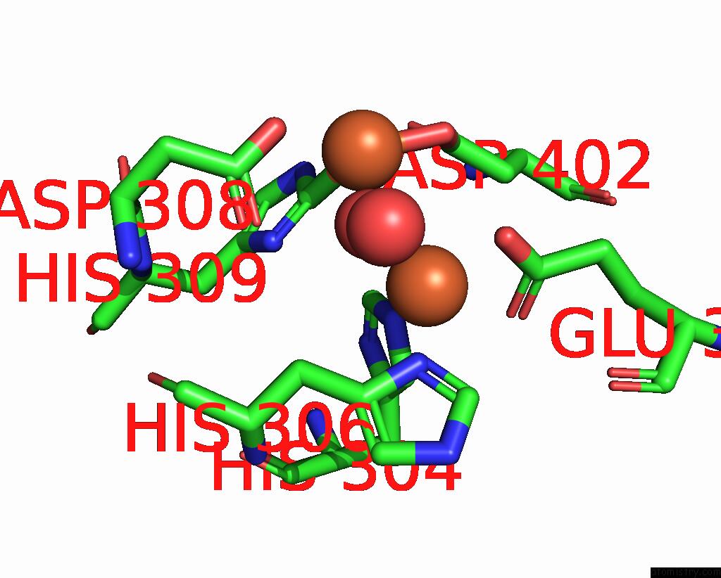
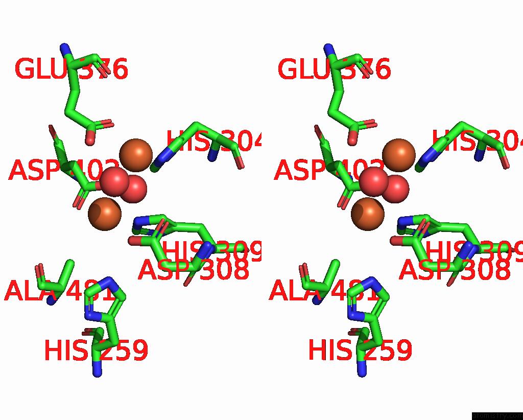
Iron binding site 1 out of 4 in 8a90

Go back to Iron Binding Sites List in 8a90
Iron binding site 1 out of 4 in the Crystal Structure of Frsh


Mono view


Stereo pair view

A full contact list of Iron with other atoms in the Fe binding site number 1 of Crystal Structure of Frsh within 5.0Å range:
probe atom residue distance (Å) B Occ
A:Fe606

b:43.9
occ:1.00
OD2 A:ASP402 1.9 77.3 1.0
CE1 A:HIS309 2.0 57.2 1.0
O A:HOH705 2.2 53.8 1.0
OD2 A:ASP308 2.3 58.1 1.0
CG A:ASP402 2.6 60.0 1.0
NE2 A:HIS309 2.8 55.8 1.0
OD1 A:ASP402 2.8 73.2 1.0
ND1 A:HIS309 3.2 69.0 1.0
CG A:ASP308 3.2 53.9 1.0
FE A:FE607 3.4 38.5 1.0
OD1 A:ASP308 3.4 66.5 1.0
OE2 A:GLU376 3.8 54.1 1.0
NE2 A:HIS259 3.9 33.0 1.0
CB A:ASP402 3.9 43.1 1.0
CD2 A:HIS309 4.0 47.8 1.0
NE2 A:HIS304 4.2 55.5 1.0
CG A:HIS309 4.2 48.6 1.0
CE1 A:HIS304 4.3 43.0 1.0
CE1 A:HIS259 4.4 43.4 1.0
CB A:ASP308 4.5 52.9 1.0
CD A:GLU376 4.7 42.4 1.0

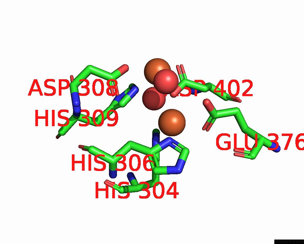
Iron binding site 2 out of 4 in 8a90

Go back to Iron Binding Sites List in 8a90
Iron binding site 2 out of 4 in the Crystal Structure of Frsh


Mono view


Stereo pair view

A full contact list of Iron with other atoms in the Fe binding site number 2 of Crystal Structure of Frsh within 5.0Å range:
probe atom residue distance (Å) B Occ
A:Fe607

b:38.5
occ:1.00
ND1 A:HIS306 2.1 46.6 1.0
NE2 A:HIS304 2.1 55.5 1.0
OE2 A:GLU376 2.1 54.1 1.0
O A:HOH705 2.1 53.8 1.0
OE1 A:GLU376 2.3 39.2 1.0
CD A:GLU376 2.5 42.4 1.0
CD2 A:HIS304 2.9 49.7 1.0
CE1 A:HIS306 3.0 58.6 1.0
CG A:HIS306 3.1 44.4 1.0
CE1 A:HIS304 3.2 43.0 1.0
FE A:FE606 3.4 43.9 1.0
CB A:HIS306 3.5 48.8 1.0
OD2 A:ASP402 3.5 77.3 1.0
CE1 A:HIS309 3.7 57.2 1.0
CG A:ASP402 3.9 60.0 1.0
CB A:ASP402 4.0 43.1 1.0
ND1 A:HIS309 4.0 69.0 1.0
CG A:GLU376 4.0 49.0 1.0
CG A:HIS304 4.1 46.2 1.0
NE2 A:HIS306 4.1 37.8 1.0
CD2 A:HIS306 4.2 35.0 1.0
ND1 A:HIS304 4.2 51.8 1.0
OD1 A:ASP308 4.3 66.5 1.0
O A:GLU376 4.6 54.4 1.0
OD1 A:ASP402 4.6 73.2 1.0
NE2 A:HIS309 4.7 55.8 1.0
CB A:GLU376 4.8 43.9 1.0
OD2 A:ASP308 4.9 58.1 1.0
O A:HOH731 4.9 64.9 1.0
CA A:HIS306 5.0 47.2 1.0

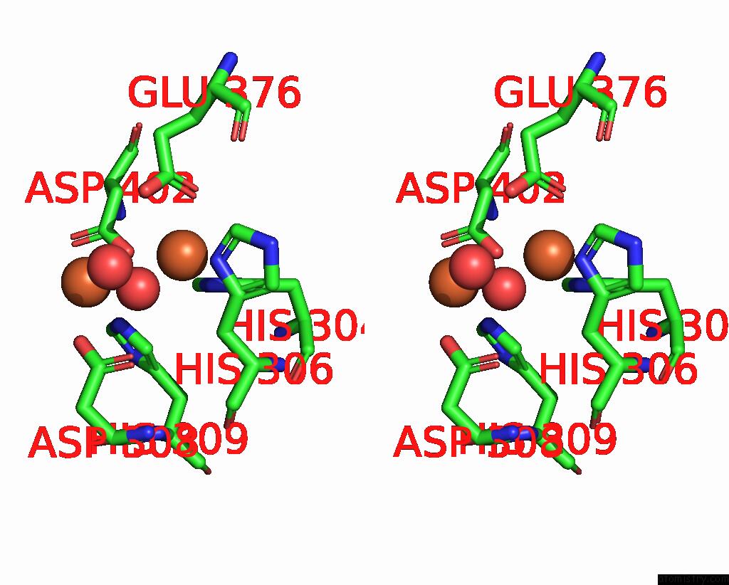
Iron binding site 3 out of 4 in 8a90

Go back to Iron Binding Sites List in 8a90
Iron binding site 3 out of 4 in the Crystal Structure of Frsh


Mono view


Stereo pair view

A full contact list of Iron with other atoms in the Fe binding site number 3 of Crystal Structure of Frsh within 5.0Å range:
probe atom residue distance (Å) B Occ
B:Fe603

b:46.1
occ:1.00
NE2 B:HIS304 2.1 38.0 1.0
O B:HOH708 2.1 71.6 1.0
ND1 B:HIS306 2.1 59.4 1.0
OE1 B:GLU376 2.4 64.9 1.0
OE2 B:GLU376 2.7 69.9 1.0
OD2 B:ASP402 2.8 58.8 1.0
CD B:GLU376 2.9 66.3 1.0
CE1 B:HIS304 2.9 41.1 1.0
CE1 B:HIS306 3.0 58.5 1.0
CG B:HIS306 3.0 56.8 1.0
CD2 B:HIS304 3.0 45.7 1.0
CB B:HIS306 3.4 52.2 1.0
FE B:FE604 3.6 51.0 1.0
CG B:ASP402 3.8 61.8 1.0
NE2 B:HIS309 4.0 52.6 1.0
CD2 B:HIS309 4.0 53.9 1.0
ND1 B:HIS304 4.0 47.7 1.0
CG B:HIS304 4.1 49.3 1.0
NE2 B:HIS306 4.1 43.3 1.0
CD2 B:HIS306 4.1 44.3 1.0
CB B:ASP402 4.3 59.1 1.0
CG B:GLU376 4.4 50.2 1.0
O B:O602 4.4 69.1 1.0
OD1 B:ASP308 4.4 73.8 1.0
O B:GLU376 4.7 68.5 1.0
OD2 B:ASP308 4.8 64.1 1.0
CA B:HIS306 4.8 63.5 1.0
OD1 B:ASP402 4.9 72.0 1.0
O B:HOH718 4.9 57.3 1.0
CE1 B:HIS309 5.0 55.2 1.0

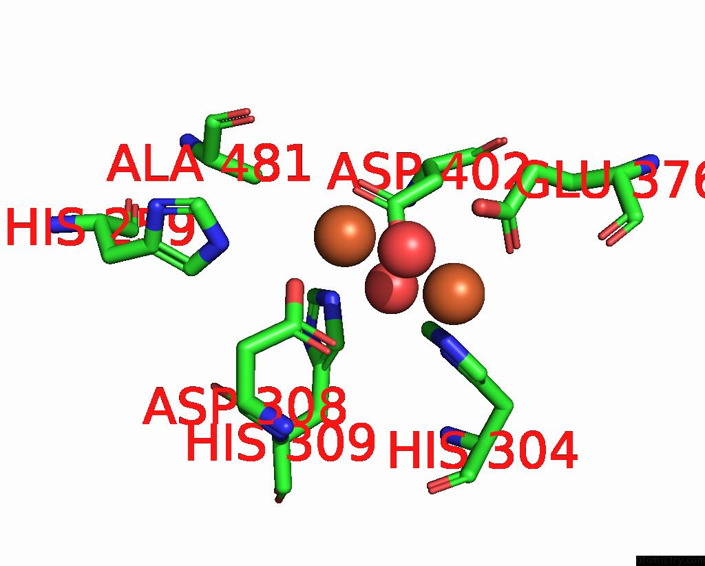
Iron binding site 4 out of 4 in 8a90

Go back to Iron Binding Sites List in 8a90
Iron binding site 4 out of 4 in the Crystal Structure of Frsh


Mono view


Stereo pair view

A full contact list of Iron with other atoms in the Fe binding site number 4 of Crystal Structure of Frsh within 5.0Å range:
probe atom residue distance (Å) B Occ
B:Fe604

b:51.0
occ:1.00
OD2 B:ASP402 2.2 58.8 1.0
O B:HOH708 2.3 71.6 1.0
NE2 B:HIS309 2.4 52.6 1.0
OD2 B:ASP308 2.5 64.1 1.0
OD1 B:ASP402 2.6 72.0 1.0
CG B:ASP402 2.7 61.8 1.0
CE1 B:HIS309 3.3 55.2 1.0
O B:O602 3.3 69.1 1.0
CD2 B:HIS309 3.4 53.9 1.0
CG B:ASP308 3.5 70.6 1.0
FE B:FE603 3.6 46.1 1.0
NE2 B:HIS259 3.7 57.6 1.0
OD1 B:ASP308 4.0 73.8 1.0
CB B:ASP402 4.0 59.1 1.0
OE2 B:GLU376 4.1 69.9 1.0
CE1 B:HIS259 4.4 64.2 1.0
ND1 B:HIS309 4.4 56.1 1.0
CG B:HIS309 4.5 40.7 1.0
CE1 B:HIS304 4.6 41.1 1.0
NE2 B:HIS304 4.6 38.0 1.0
CD2 B:HIS259 4.7 53.0 1.0
CB B:ALA481 4.8 50.3 1.0
O B:ALA481 4.8 69.3 1.0
CB B:ASP308 4.8 42.4 1.0
CD B:GLU376 4.9 66.3 1.0
O B:HOH718 5.0 57.3 1.0

Reference:

D.A.Wirtz, N.Schneberger, S.Kloppel, R.Richarz, M.Geyer, G.M.Konig, G.Hagelueken, M.Crusemann. Adenylation Domain-Guided Recruitment of Trans- Acting Nonheme Monooxygenases in Nonribosomal Peptide Biosynthesis. Acs Chem.Biol. 2023.
ISSN: ESSN 1554-8937
PubMed: 37366538
DOI: 10.1021/ACSCHEMBIO.3C00106
Page generated: Thu Jul 27 15:55:55 2023

Last articles

Zn in 8WB0
Zn in 8WAX
Zn in 8WAU
Zn in 8WAZ
Zn in 8WAY
Zn in 8WAV
Zn in 8WAW
Zn in 8WAT
Zn in 8W7M
Zn in 8WD3
© Copyright 2008-2020 by atomistry.com
Home   |    Site Map   |    Copyright   |    Contact us   |    Privacy