Iron »
PDB 8bgw-8ca3 »
8c7c »
Iron in PDB 8c7c: Double Mutant V(M84)C/A(L278)C Structure of Photosynthetic Reaction Center From Cereibacter Sphaeroides Strain Rv
Protein crystallography data
The structure of Double Mutant V(M84)C/A(L278)C Structure of Photosynthetic Reaction Center From Cereibacter Sphaeroides Strain Rv, PDB code: 8c7c
was solved by
A.Gabdulkhakov,
G.Selikhanov,
T.Fufina,
L.Vasilieva,
A.Atamas,
D.Uhimchuk,
with X-Ray Crystallography technique. A brief refinement statistics is given in the table below:
| Resolution Low / High (Å) | 41.99 / 2.60 |
| Space group | C 1 2 1 |
| Cell size a, b, c (Å), α, β, γ (°) | 253.13, 75.98, 65.85, 90, 95.58, 90 |
| R / Rfree (%) | 19.3 / 24.9 |
Other elements in 8c7c:
The structure of Double Mutant V(M84)C/A(L278)C Structure of Photosynthetic Reaction Center From Cereibacter Sphaeroides Strain Rv also contains other interesting chemical elements:
| Magnesium | (Mg) | 4 atoms |
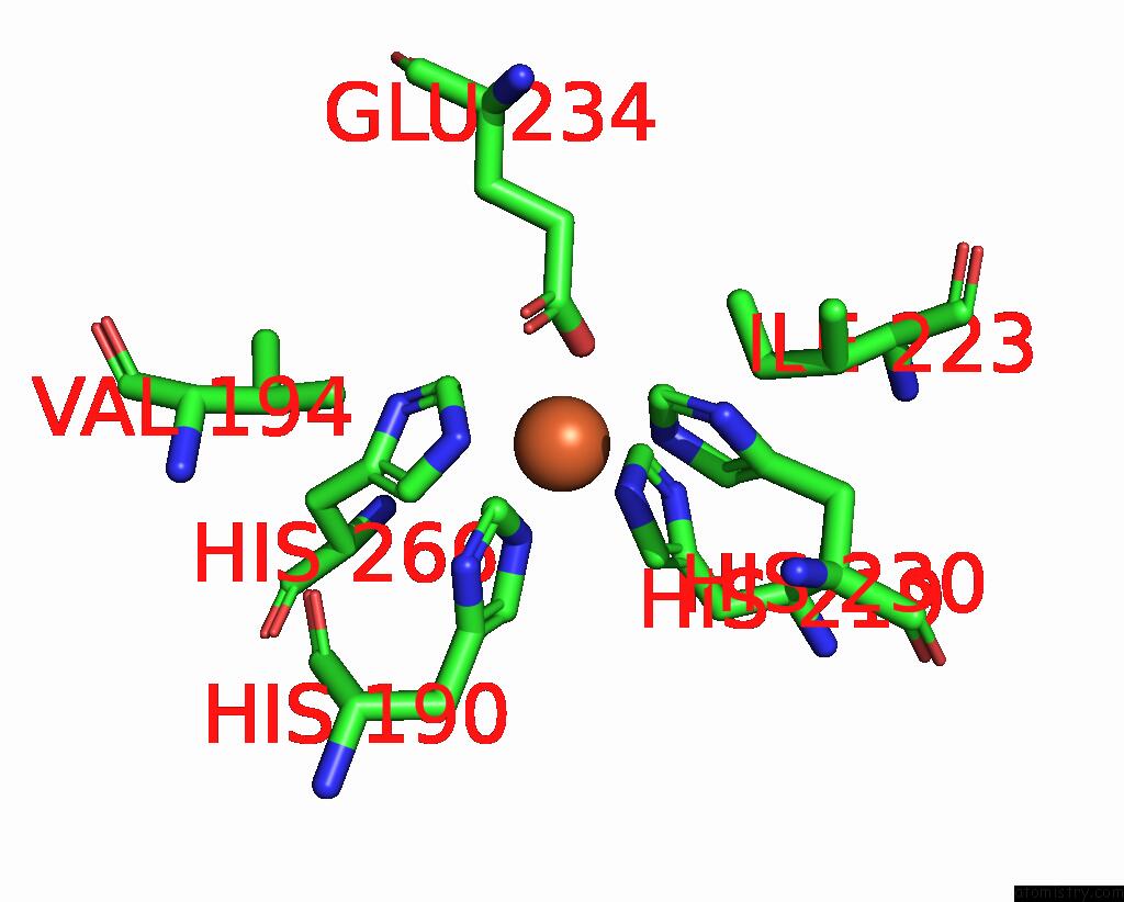
Iron Binding Sites:
The binding sites of Iron atom in the Double Mutant V(M84)C/A(L278)C Structure of Photosynthetic Reaction Center From Cereibacter Sphaeroides Strain Rv
(pdb code 8c7c). This binding sites where shown within
5.0 Angstroms radius around Iron atom.
In total only one binding site of Iron was determined in the Double Mutant V(M84)C/A(L278)C Structure of Photosynthetic Reaction Center From Cereibacter Sphaeroides Strain Rv, PDB code: 8c7c:
In total only one binding site of Iron was determined in the Double Mutant V(M84)C/A(L278)C Structure of Photosynthetic Reaction Center From Cereibacter Sphaeroides Strain Rv, PDB code: 8c7c:
Iron binding site 1 out of 1 in 8c7c
Go back to
Iron binding site 1 out
of 1 in the Double Mutant V(M84)C/A(L278)C Structure of Photosynthetic Reaction Center From Cereibacter Sphaeroides Strain Rv
Mono view
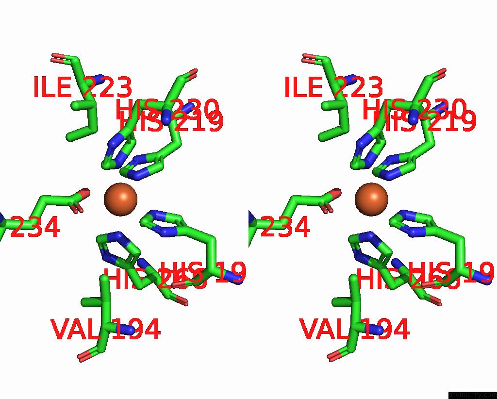
Stereo pair view
Mono view
Stereo pair view
A full contact list of Iron with other atoms in the Fe binding
site number 1 of Double Mutant V(M84)C/A(L278)C Structure of Photosynthetic Reaction Center From Cereibacter Sphaeroides Strain Rv within 5.0Å range:
|
Reference:
G.Selikhanov,
A.Atamas,
D.Yukhimchuk,
T.Fufina,
L.Vasilieva,
A.Gabdulkhakov.
Stabilization of Cereibacter Sphaeroides Photosynthetic Reaction Center By the Introduction of Disulfide Bonds. Membranes (Basel) V. 13 2023.
ISSN: ESSN 2077-0375
PubMed: 36837657
DOI: 10.3390/MEMBRANES13020154
Page generated: Fri Aug 9 20:30:16 2024
ISSN: ESSN 2077-0375
PubMed: 36837657
DOI: 10.3390/MEMBRANES13020154
Last articles
Zn in 9J0NZn in 9J0O
Zn in 9J0P
Zn in 9FJX
Zn in 9EKB
Zn in 9C0F
Zn in 9CAH
Zn in 9CH0
Zn in 9CH3
Zn in 9CH1