Iron »
PDB 8cah-8csr »
8cjo »
Iron in PDB 8cjo: Crystal Structure of Human Tryptophan Hydroxylase 1 in Complex with Inhibitor Km-06-004
Enzymatic activity of Crystal Structure of Human Tryptophan Hydroxylase 1 in Complex with Inhibitor Km-06-004
All present enzymatic activity of Crystal Structure of Human Tryptophan Hydroxylase 1 in Complex with Inhibitor Km-06-004:
1.14.16.4;
1.14.16.4;
Protein crystallography data
The structure of Crystal Structure of Human Tryptophan Hydroxylase 1 in Complex with Inhibitor Km-06-004, PDB code: 8cjo
was solved by
A.Schuetz,
K.Mallow,
M.Nazare,
E.Specker,
U.Heinemann,
with X-Ray Crystallography technique. A brief refinement statistics is given in the table below:
| Resolution Low / High (Å) | 44.31 / 1.87 |
| Space group | P 1 21 1 |
| Cell size a, b, c (Å), α, β, γ (°) | 47.218, 57.776, 69.6, 90, 109.35, 90 |
| R / Rfree (%) | 16.9 / 20.9 |
Other elements in 8cjo:
The structure of Crystal Structure of Human Tryptophan Hydroxylase 1 in Complex with Inhibitor Km-06-004 also contains other interesting chemical elements:
| Fluorine | (F) | 1 atom |
| Chlorine | (Cl) | 1 atom |
Iron Binding Sites:
The binding sites of Iron atom in the Crystal Structure of Human Tryptophan Hydroxylase 1 in Complex with Inhibitor Km-06-004
(pdb code 8cjo). This binding sites where shown within
5.0 Angstroms radius around Iron atom.
In total only one binding site of Iron was determined in the Crystal Structure of Human Tryptophan Hydroxylase 1 in Complex with Inhibitor Km-06-004, PDB code: 8cjo:
In total only one binding site of Iron was determined in the Crystal Structure of Human Tryptophan Hydroxylase 1 in Complex with Inhibitor Km-06-004, PDB code: 8cjo:
Iron binding site 1 out of 1 in 8cjo
Go back to
Iron binding site 1 out
of 1 in the Crystal Structure of Human Tryptophan Hydroxylase 1 in Complex with Inhibitor Km-06-004
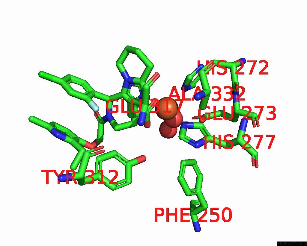
Mono view
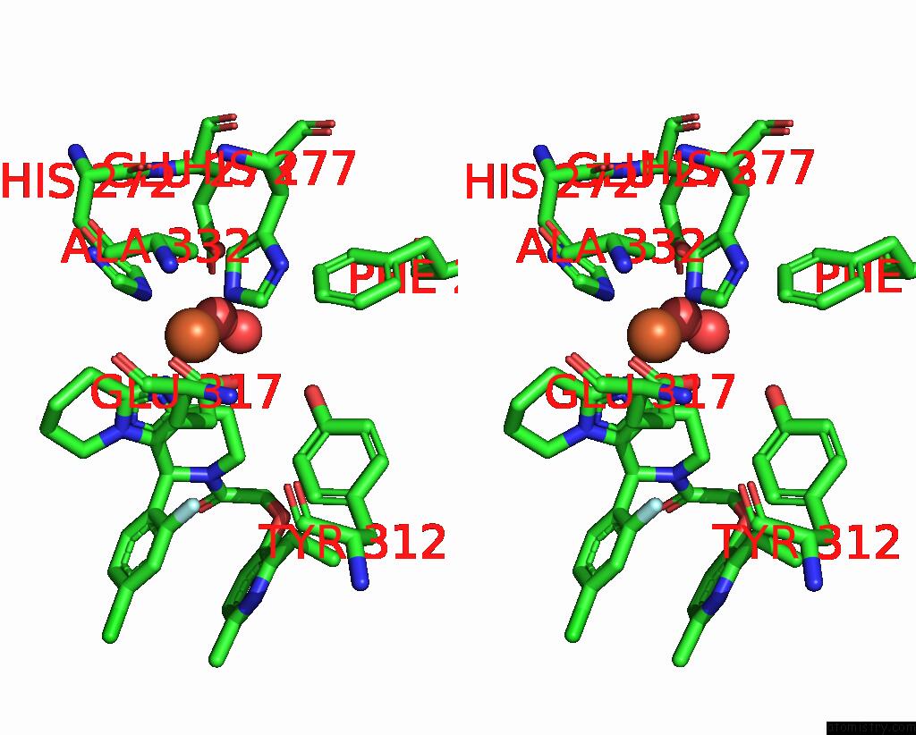
Stereo pair view
Mono view
Stereo pair view
A full contact list of Iron with other atoms in the Fe binding
site number 1 of Crystal Structure of Human Tryptophan Hydroxylase 1 in Complex with Inhibitor Km-06-004 within 5.0Å range:
|
Reference:
E.Specker,
R.Wesolowski,
A.Schutz,
S.Matthes,
K.Mallow,
M.Wasinska-Kalwa,
L.Winkler,
A.Oder,
N.Alenina,
D.Pleimes,
J.P.Von Kries,
U.Heinemann,
M.Bader,
M.Nazare.
Structure-Based Design of Xanthine-Imidazopyridines and -Imidazothiazoles As Highly Potent and in Vivo Efficacious Tryptophan Hydroxylase Inhibitors. J.Med.Chem. V. 66 14866 2023.
ISSN: ISSN 0022-2623
PubMed: 37905925
DOI: 10.1021/ACS.JMEDCHEM.3C01454
Page generated: Thu Aug 7 14:44:49 2025
ISSN: ISSN 0022-2623
PubMed: 37905925
DOI: 10.1021/ACS.JMEDCHEM.3C01454
Last articles
Mg in 2OQYMg in 2OUP
Mg in 2OUN
Mg in 2OU7
Mg in 2OTG
Mg in 2OSB
Mg in 2OS8
Mg in 2ORI
Mg in 2ORW
Mg in 2ONP