Iron »
PDB 8djs-8e3t »
8drj »
Iron in PDB 8drj: Apo B2 Dimer (H60/H100/H104) Formed in the Presence of Cu(II)
Protein crystallography data
The structure of Apo B2 Dimer (H60/H100/H104) Formed in the Presence of Cu(II), PDB code: 8drj
was solved by
T.S.Choi,
F.A.Tezcan,
with X-Ray Crystallography technique. A brief refinement statistics is given in the table below:
| Resolution Low / High (Å) | 44.86 / 2.40 |
| Space group | C 1 2 1 |
| Cell size a, b, c (Å), α, β, γ (°) | 89.32, 48.02, 45.12, 90, 96.18, 90 |
| R / Rfree (%) | 19.1 / 28.3 |
Iron Binding Sites:
The binding sites of Iron atom in the Apo B2 Dimer (H60/H100/H104) Formed in the Presence of Cu(II)
(pdb code 8drj). This binding sites where shown within
5.0 Angstroms radius around Iron atom.
In total 2 binding sites of Iron where determined in the Apo B2 Dimer (H60/H100/H104) Formed in the Presence of Cu(II), PDB code: 8drj:
Jump to Iron binding site number: 1; 2;
In total 2 binding sites of Iron where determined in the Apo B2 Dimer (H60/H100/H104) Formed in the Presence of Cu(II), PDB code: 8drj:
Jump to Iron binding site number: 1; 2;
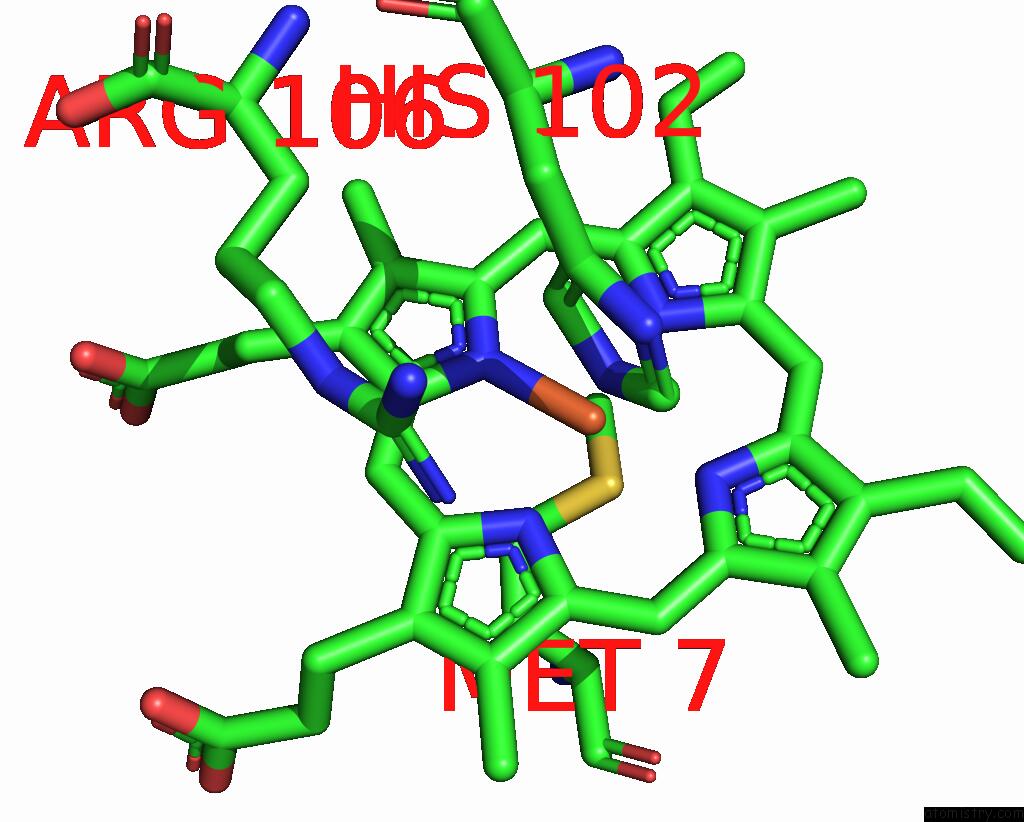
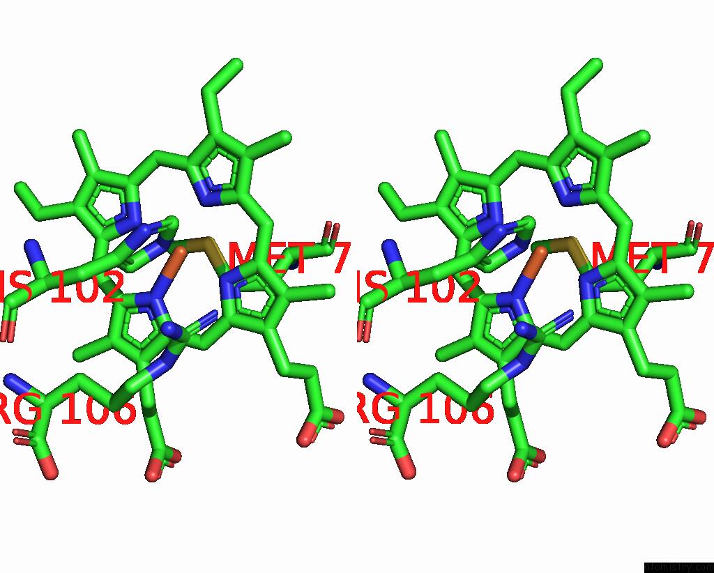
Iron binding site 1 out of 2 in 8drj
Go back to
Iron binding site 1 out
of 2 in the Apo B2 Dimer (H60/H100/H104) Formed in the Presence of Cu(II)

Mono view

Stereo pair view
Mono view
Stereo pair view
A full contact list of Iron with other atoms in the Fe binding
site number 1 of Apo B2 Dimer (H60/H100/H104) Formed in the Presence of Cu(II) within 5.0Å range:
|
Iron binding site 2 out of 2 in 8drj
Go back to
Iron binding site 2 out
of 2 in the Apo B2 Dimer (H60/H100/H104) Formed in the Presence of Cu(II)
Mono view
Stereo pair view
Mono view
Stereo pair view
A full contact list of Iron with other atoms in the Fe binding
site number 2 of Apo B2 Dimer (H60/H100/H104) Formed in the Presence of Cu(II) within 5.0Å range:
|
Reference:
T.S.Choi,
F.A.Tezcan.
Design of A Flexible, Zn-Selective Protein Scaffold That Displays Anti-Irving-Williams Behavior. J.Am.Chem.Soc. V. 144 18090 2022.
ISSN: ESSN 1520-5126
PubMed: 36154053
DOI: 10.1021/JACS.2C08050
Page generated: Sat Aug 10 00:17:09 2024
ISSN: ESSN 1520-5126
PubMed: 36154053
DOI: 10.1021/JACS.2C08050
Last articles
Zn in 9MJ5Zn in 9HNW
Zn in 9G0L
Zn in 9FNE
Zn in 9DZN
Zn in 9E0I
Zn in 9D32
Zn in 9DAK
Zn in 8ZXC
Zn in 8ZUF