Iron »
PDB 8djs-8e3t »
8dwf »
Iron in PDB 8dwf: Glycosylase Muty Variant E43S in Complex with Dna Containing D(8-Oxo- G) Paired with Substrate Adenine
Enzymatic activity of Glycosylase Muty Variant E43S in Complex with Dna Containing D(8-Oxo- G) Paired with Substrate Adenine
All present enzymatic activity of Glycosylase Muty Variant E43S in Complex with Dna Containing D(8-Oxo- G) Paired with Substrate Adenine:
3.2.2.31;
3.2.2.31;
Protein crystallography data
The structure of Glycosylase Muty Variant E43S in Complex with Dna Containing D(8-Oxo- G) Paired with Substrate Adenine, PDB code: 8dwf
was solved by
L.P.Russelburg,
M.Demir,
S.S.David,
M.P.Horvath,
with X-Ray Crystallography technique. A brief refinement statistics is given in the table below:
| Resolution Low / High (Å) | 73.00 / 2.60 |
| Space group | P 1 21 1 |
| Cell size a, b, c (Å), α, β, γ (°) | 49.08, 137.9, 74.52, 90, 101.44, 90 |
| R / Rfree (%) | 20.7 / 25.3 |
Other elements in 8dwf:
The structure of Glycosylase Muty Variant E43S in Complex with Dna Containing D(8-Oxo- G) Paired with Substrate Adenine also contains other interesting chemical elements:
| Calcium | (Ca) | 4 atoms |
Iron Binding Sites:
The binding sites of Iron atom in the Glycosylase Muty Variant E43S in Complex with Dna Containing D(8-Oxo- G) Paired with Substrate Adenine
(pdb code 8dwf). This binding sites where shown within
5.0 Angstroms radius around Iron atom.
In total 8 binding sites of Iron where determined in the Glycosylase Muty Variant E43S in Complex with Dna Containing D(8-Oxo- G) Paired with Substrate Adenine, PDB code: 8dwf:
Jump to Iron binding site number: 1; 2; 3; 4; 5; 6; 7; 8;
In total 8 binding sites of Iron where determined in the Glycosylase Muty Variant E43S in Complex with Dna Containing D(8-Oxo- G) Paired with Substrate Adenine, PDB code: 8dwf:
Jump to Iron binding site number: 1; 2; 3; 4; 5; 6; 7; 8;
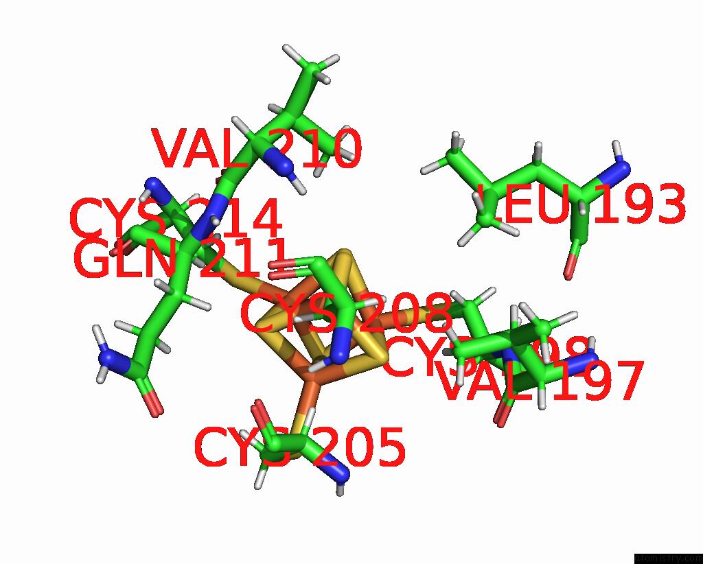
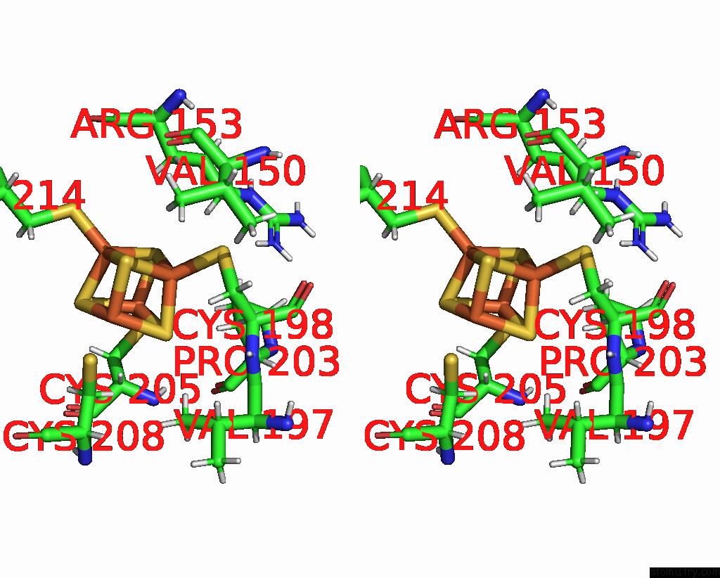
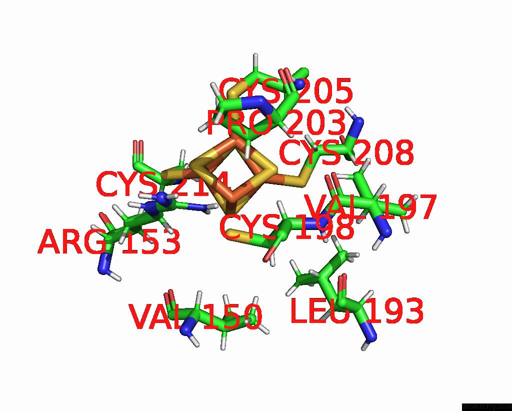
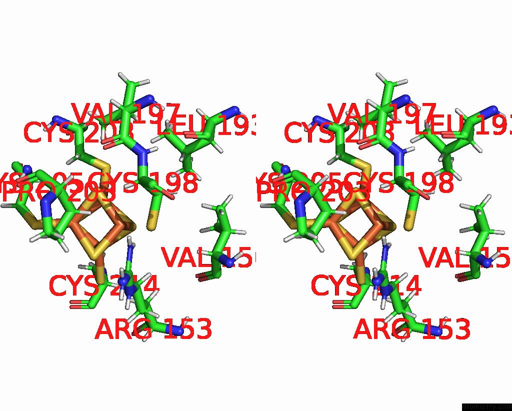
Iron binding site 1 out of 8 in 8dwf
Go back to
Iron binding site 1 out
of 8 in the Glycosylase Muty Variant E43S in Complex with Dna Containing D(8-Oxo- G) Paired with Substrate Adenine
Mono view
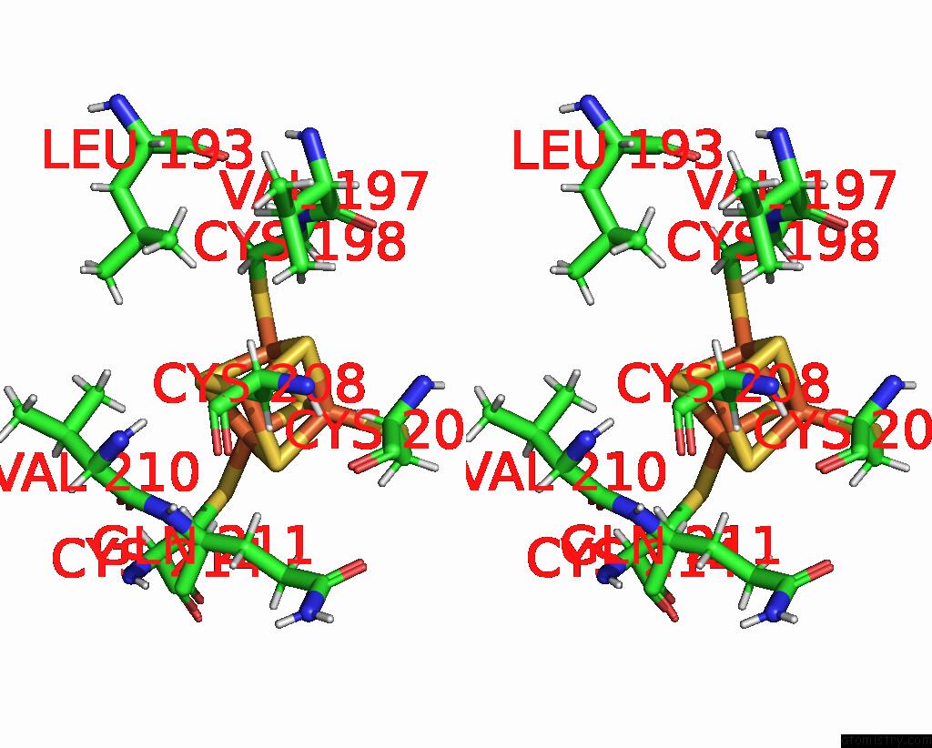
Stereo pair view
Mono view
Stereo pair view
A full contact list of Iron with other atoms in the Fe binding
site number 1 of Glycosylase Muty Variant E43S in Complex with Dna Containing D(8-Oxo- G) Paired with Substrate Adenine within 5.0Å range:
|
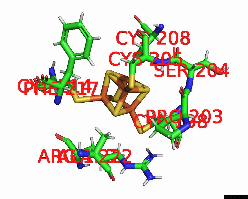
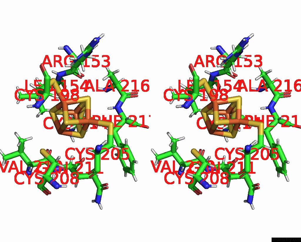
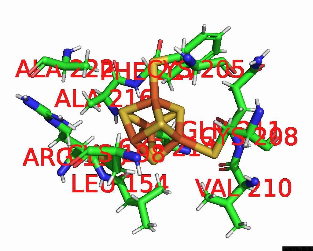
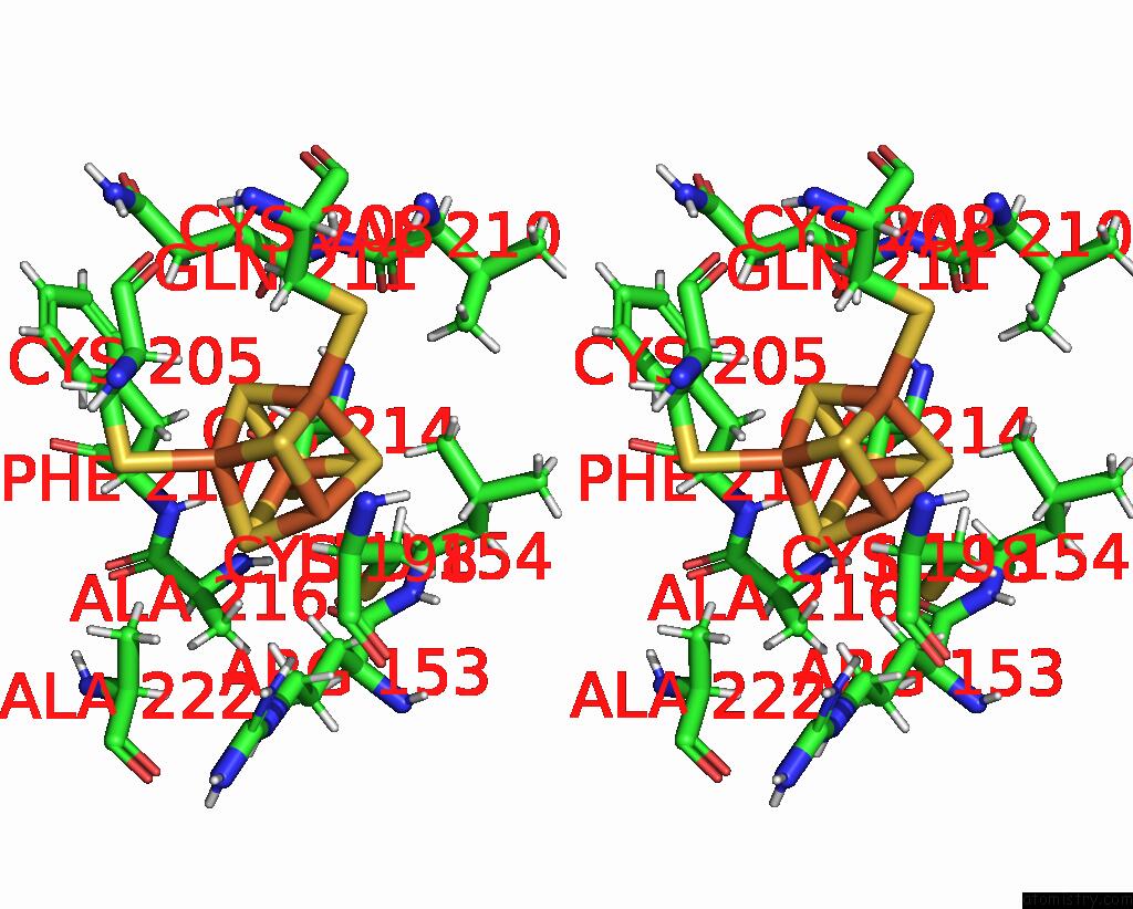
Iron binding site 2 out of 8 in 8dwf
Go back to
Iron binding site 2 out
of 8 in the Glycosylase Muty Variant E43S in Complex with Dna Containing D(8-Oxo- G) Paired with Substrate Adenine
Mono view
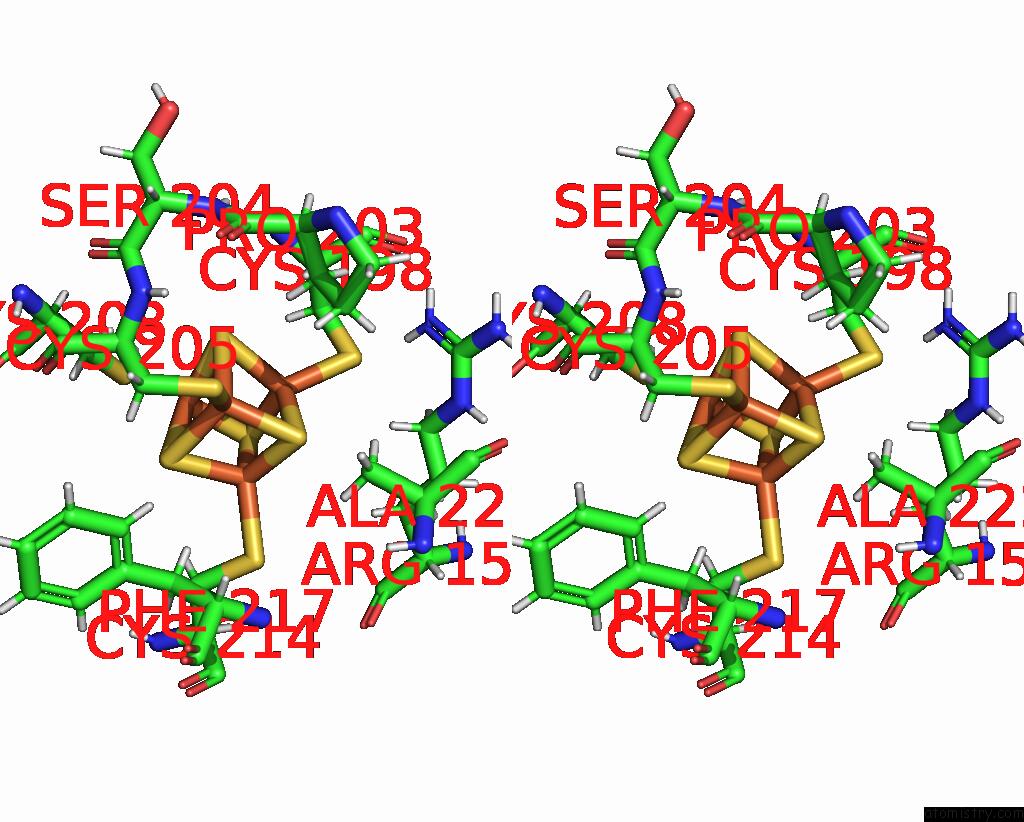
Stereo pair view
Mono view
Stereo pair view
A full contact list of Iron with other atoms in the Fe binding
site number 2 of Glycosylase Muty Variant E43S in Complex with Dna Containing D(8-Oxo- G) Paired with Substrate Adenine within 5.0Å range:
|
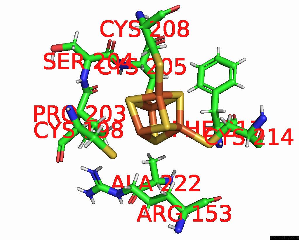
Iron binding site 3 out of 8 in 8dwf
Go back to
Iron binding site 3 out
of 8 in the Glycosylase Muty Variant E43S in Complex with Dna Containing D(8-Oxo- G) Paired with Substrate Adenine

Mono view
Stereo pair view
Mono view
Stereo pair view
A full contact list of Iron with other atoms in the Fe binding
site number 3 of Glycosylase Muty Variant E43S in Complex with Dna Containing D(8-Oxo- G) Paired with Substrate Adenine within 5.0Å range:
|
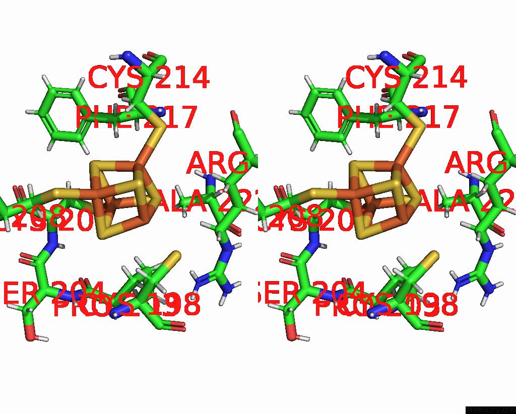
Iron binding site 4 out of 8 in 8dwf
Go back to
Iron binding site 4 out
of 8 in the Glycosylase Muty Variant E43S in Complex with Dna Containing D(8-Oxo- G) Paired with Substrate Adenine

Mono view
Stereo pair view
Mono view
Stereo pair view
A full contact list of Iron with other atoms in the Fe binding
site number 4 of Glycosylase Muty Variant E43S in Complex with Dna Containing D(8-Oxo- G) Paired with Substrate Adenine within 5.0Å range:
|
Iron binding site 5 out of 8 in 8dwf
Go back to
Iron binding site 5 out
of 8 in the Glycosylase Muty Variant E43S in Complex with Dna Containing D(8-Oxo- G) Paired with Substrate Adenine

Mono view

Stereo pair view
Mono view
Stereo pair view
A full contact list of Iron with other atoms in the Fe binding
site number 5 of Glycosylase Muty Variant E43S in Complex with Dna Containing D(8-Oxo- G) Paired with Substrate Adenine within 5.0Å range:
|
Iron binding site 6 out of 8 in 8dwf
Go back to
Iron binding site 6 out
of 8 in the Glycosylase Muty Variant E43S in Complex with Dna Containing D(8-Oxo- G) Paired with Substrate Adenine
Mono view
Stereo pair view
Mono view
Stereo pair view
A full contact list of Iron with other atoms in the Fe binding
site number 6 of Glycosylase Muty Variant E43S in Complex with Dna Containing D(8-Oxo- G) Paired with Substrate Adenine within 5.0Å range:
|
Iron binding site 7 out of 8 in 8dwf
Go back to
Iron binding site 7 out
of 8 in the Glycosylase Muty Variant E43S in Complex with Dna Containing D(8-Oxo- G) Paired with Substrate Adenine
Mono view
Stereo pair view
Mono view
Stereo pair view
A full contact list of Iron with other atoms in the Fe binding
site number 7 of Glycosylase Muty Variant E43S in Complex with Dna Containing D(8-Oxo- G) Paired with Substrate Adenine within 5.0Å range:
|
Iron binding site 8 out of 8 in 8dwf
Go back to
Iron binding site 8 out
of 8 in the Glycosylase Muty Variant E43S in Complex with Dna Containing D(8-Oxo- G) Paired with Substrate Adenine
Mono view
Stereo pair view
Mono view
Stereo pair view
A full contact list of Iron with other atoms in the Fe binding
site number 8 of Glycosylase Muty Variant E43S in Complex with Dna Containing D(8-Oxo- G) Paired with Substrate Adenine within 5.0Å range:
|
Reference:
L.P.Russelburg,
M.Demir,
K.Cedeno,
S.S.David,
M.P.Horvath.
Structural Basis For Base Engagement and Stereochemistry Revealed By Alteration of Catalytic Residue GLU43 in Dna Repair Glycosylase Muty To Be Published.
Page generated: Sat Aug 10 00:27:54 2024
Last articles
Zn in 9JYWZn in 9IR4
Zn in 9IR3
Zn in 9GMX
Zn in 9GMW
Zn in 9JEJ
Zn in 9ERF
Zn in 9ERE
Zn in 9EGV
Zn in 9EGW