Iron »
PDB 8e3u-8eqm »
8e7w »
Iron in PDB 8e7w: Rstspo A139T with Heme
Protein crystallography data
The structure of Rstspo A139T with Heme, PDB code: 8e7w
was solved by
J.Liu,
C.Hiser,
F.Li,
R.Garavito,
S.Ferguson-Miller,
with X-Ray Crystallography technique. A brief refinement statistics is given in the table below:
| Resolution Low / High (Å) | 28.95 / 2.10 |
| Space group | C 1 2 1 |
| Cell size a, b, c (Å), α, β, γ (°) | 58.399, 99.568, 95.812, 90, 100.16, 90 |
| R / Rfree (%) | 19.5 / 25.3 |
Iron Binding Sites:
The binding sites of Iron atom in the Rstspo A139T with Heme
(pdb code 8e7w). This binding sites where shown within
5.0 Angstroms radius around Iron atom.
In total only one binding site of Iron was determined in the Rstspo A139T with Heme, PDB code: 8e7w:
In total only one binding site of Iron was determined in the Rstspo A139T with Heme, PDB code: 8e7w:
Iron binding site 1 out of 1 in 8e7w
Go back to
Iron binding site 1 out
of 1 in the Rstspo A139T with Heme

Mono view
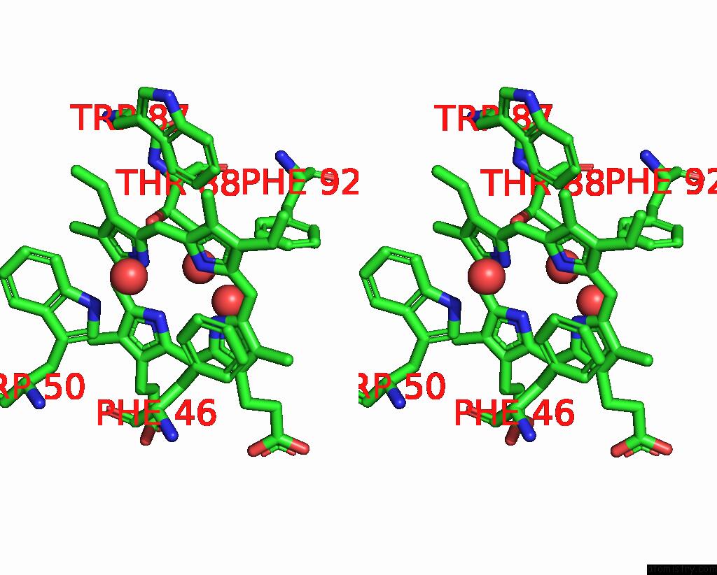
Stereo pair view
Mono view
Stereo pair view
A full contact list of Iron with other atoms in the Fe binding
site number 1 of Rstspo A139T with Heme within 5.0Å range:
|
Reference:
J.Liu,
C.Hiser,
F.Li,
R.Hall,
R.M.Garavito,
S.Ferguson-Miller.
New Tspo Crystal Structures of Mutant and Heme-Bound Forms with Altered Flexibility, Ligand Binding, and Porphyrin Degradation Activity. Biochemistry 2023.
ISSN: ISSN 0006-2960
PubMed: 36947867
DOI: 10.1021/ACS.BIOCHEM.2C00612
Page generated: Sat Aug 10 00:54:06 2024
ISSN: ISSN 0006-2960
PubMed: 36947867
DOI: 10.1021/ACS.BIOCHEM.2C00612
Last articles
Zn in 9MJ5Zn in 9HNW
Zn in 9G0L
Zn in 9FNE
Zn in 9DZN
Zn in 9E0I
Zn in 9D32
Zn in 9DAK
Zn in 8ZXC
Zn in 8ZUF