Iron »
PDB 8e3u-8eqm »
8e83 »
Iron in PDB 8e83: Structure of 2-Hydroxyisoflavanone Synthase From Medicago Truncatula
Enzymatic activity of Structure of 2-Hydroxyisoflavanone Synthase From Medicago Truncatula
All present enzymatic activity of Structure of 2-Hydroxyisoflavanone Synthase From Medicago Truncatula:
1.14.14.87;
1.14.14.87;
Protein crystallography data
The structure of Structure of 2-Hydroxyisoflavanone Synthase From Medicago Truncatula, PDB code: 8e83
was solved by
H.Pan,
X.Wang,
with X-Ray Crystallography technique. A brief refinement statistics is given in the table below:
| Resolution Low / High (Å) | 42.70 / 2.00 |
| Space group | P 1 21 1 |
| Cell size a, b, c (Å), α, β, γ (°) | 50.119, 73.787, 148.671, 90, 93.48, 90 |
| R / Rfree (%) | 19.2 / 22.8 |
Iron Binding Sites:
The binding sites of Iron atom in the Structure of 2-Hydroxyisoflavanone Synthase From Medicago Truncatula
(pdb code 8e83). This binding sites where shown within
5.0 Angstroms radius around Iron atom.
In total 2 binding sites of Iron where determined in the Structure of 2-Hydroxyisoflavanone Synthase From Medicago Truncatula, PDB code: 8e83:
Jump to Iron binding site number: 1; 2;
In total 2 binding sites of Iron where determined in the Structure of 2-Hydroxyisoflavanone Synthase From Medicago Truncatula, PDB code: 8e83:
Jump to Iron binding site number: 1; 2;
Iron binding site 1 out of 2 in 8e83
Go back to
Iron binding site 1 out
of 2 in the Structure of 2-Hydroxyisoflavanone Synthase From Medicago Truncatula
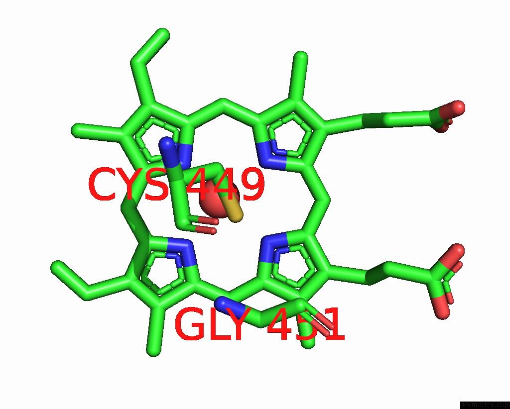
Mono view
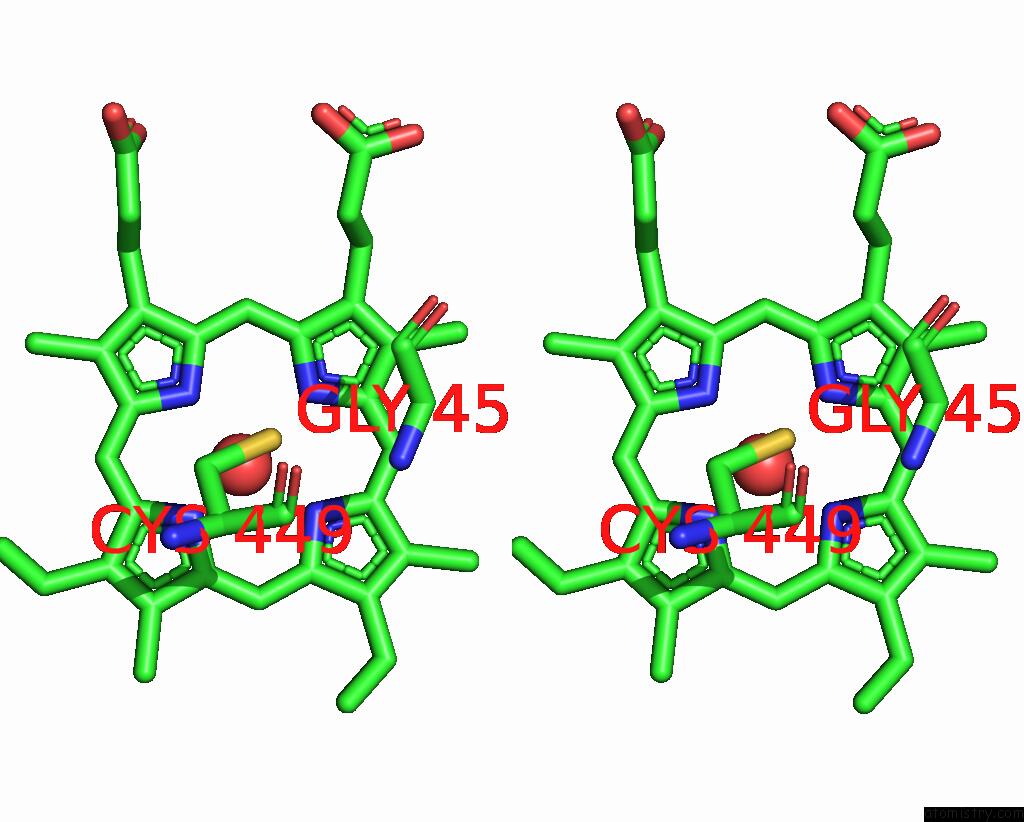
Stereo pair view
Mono view
Stereo pair view
A full contact list of Iron with other atoms in the Fe binding
site number 1 of Structure of 2-Hydroxyisoflavanone Synthase From Medicago Truncatula within 5.0Å range:
|
Iron binding site 2 out of 2 in 8e83
Go back to
Iron binding site 2 out
of 2 in the Structure of 2-Hydroxyisoflavanone Synthase From Medicago Truncatula
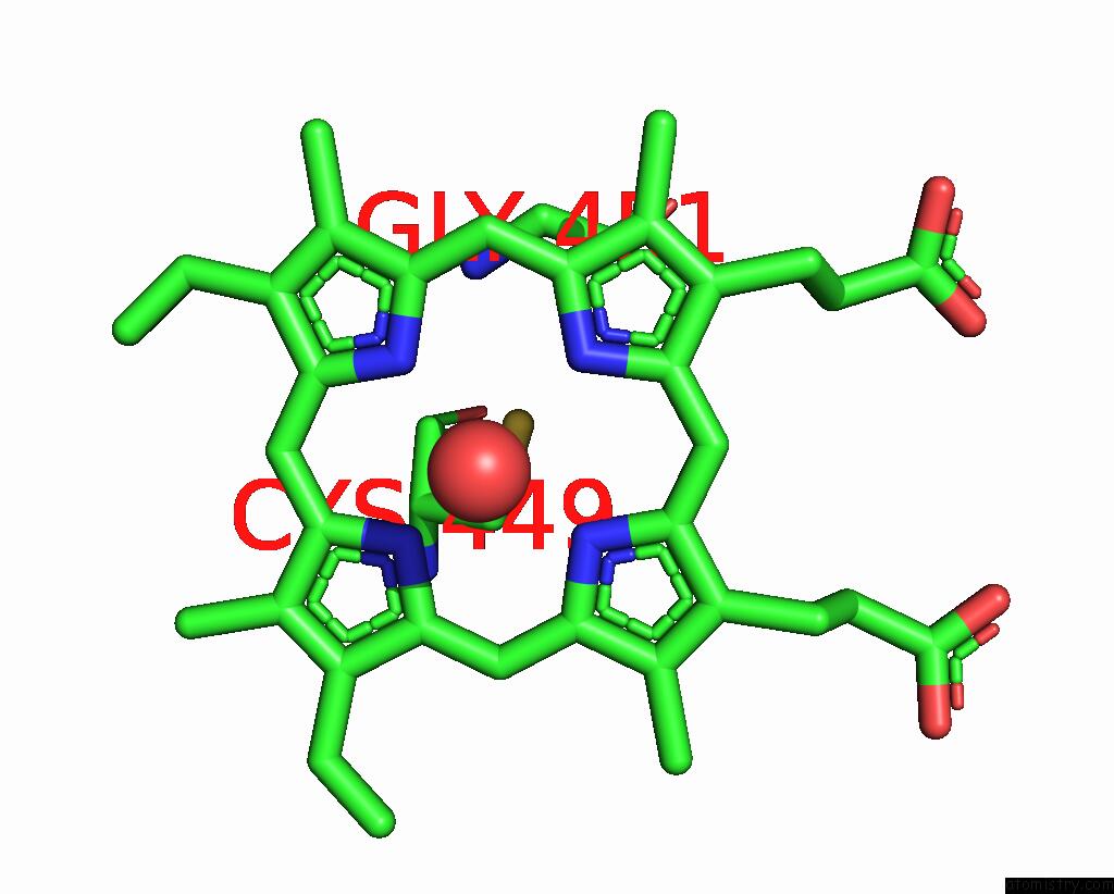
Mono view
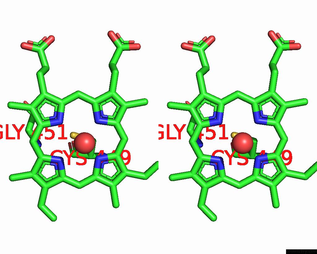
Stereo pair view
Mono view
Stereo pair view
A full contact list of Iron with other atoms in the Fe binding
site number 2 of Structure of 2-Hydroxyisoflavanone Synthase From Medicago Truncatula within 5.0Å range:
|
Reference:
X.Wang,
H.Pan,
S.Sagurthi,
V.Paris,
C.Zhuo,
R.A.Dixon.
The Protein Conformational Basis of Isoflavone Biosynthesis. Commun Biol V. 5 1249 2022.
ISSN: ESSN 2399-3642
PubMed: 36376429
DOI: 10.1038/S42003-022-04222-X
Page generated: Sat Aug 10 00:55:47 2024
ISSN: ESSN 2399-3642
PubMed: 36376429
DOI: 10.1038/S42003-022-04222-X
Last articles
Zn in 9J0NZn in 9J0O
Zn in 9J0P
Zn in 9FJX
Zn in 9EKB
Zn in 9C0F
Zn in 9CAH
Zn in 9CH0
Zn in 9CH3
Zn in 9CH1