Iron »
PDB 8e3u-8eqm »
8ed4 »
Iron in PDB 8ed4: Structure of the Complex Between the Arsenite Oxidase and Its Native Electron Acceptor Cytochrome C552 From Pseudorhizobium Sp. Str. Nt-26
Enzymatic activity of Structure of the Complex Between the Arsenite Oxidase and Its Native Electron Acceptor Cytochrome C552 From Pseudorhizobium Sp. Str. Nt-26
All present enzymatic activity of Structure of the Complex Between the Arsenite Oxidase and Its Native Electron Acceptor Cytochrome C552 From Pseudorhizobium Sp. Str. Nt-26:
1.20.98.1;
1.20.98.1;
Protein crystallography data
The structure of Structure of the Complex Between the Arsenite Oxidase and Its Native Electron Acceptor Cytochrome C552 From Pseudorhizobium Sp. Str. Nt-26, PDB code: 8ed4
was solved by
M.J.Maher,
N.Poddar,
with X-Ray Crystallography technique. A brief refinement statistics is given in the table below:
| Resolution Low / High (Å) | 49.23 / 2.25 |
| Space group | P 1 21 1 |
| Cell size a, b, c (Å), α, β, γ (°) | 129.4, 126.56, 148.02, 90, 107.81, 90 |
| R / Rfree (%) | 18.1 / 23 |
Other elements in 8ed4:
The structure of Structure of the Complex Between the Arsenite Oxidase and Its Native Electron Acceptor Cytochrome C552 From Pseudorhizobium Sp. Str. Nt-26 also contains other interesting chemical elements:
| Molybdenum | (Mo) | 4 atoms |
Iron Binding Sites:
Pages:
>>> Page 1 <<< Page 2, Binding sites: 11 - 20; Page 3, Binding sites: 21 - 24;Binding sites:
The binding sites of Iron atom in the Structure of the Complex Between the Arsenite Oxidase and Its Native Electron Acceptor Cytochrome C552 From Pseudorhizobium Sp. Str. Nt-26 (pdb code 8ed4). This binding sites where shown within 5.0 Angstroms radius around Iron atom.In total 24 binding sites of Iron where determined in the Structure of the Complex Between the Arsenite Oxidase and Its Native Electron Acceptor Cytochrome C552 From Pseudorhizobium Sp. Str. Nt-26, PDB code: 8ed4:
Jump to Iron binding site number: 1; 2; 3; 4; 5; 6; 7; 8; 9; 10;
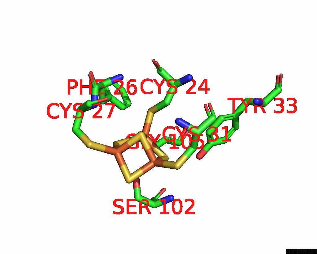
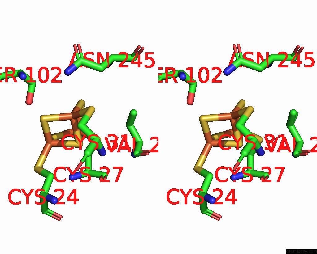
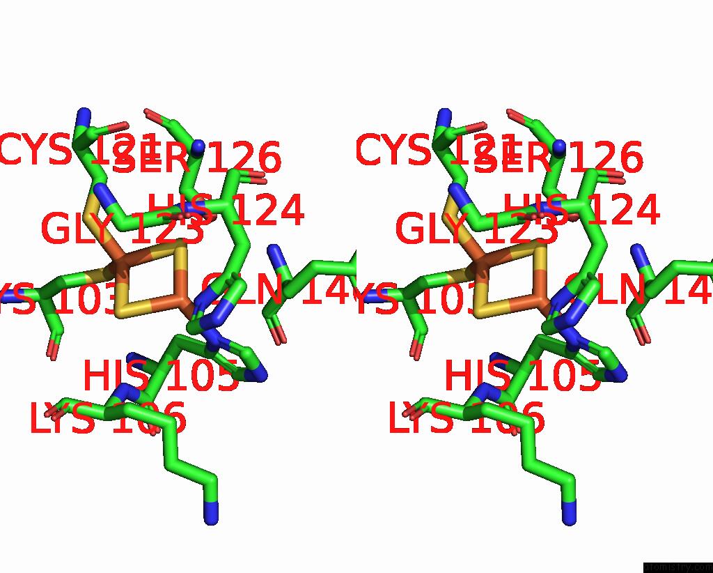
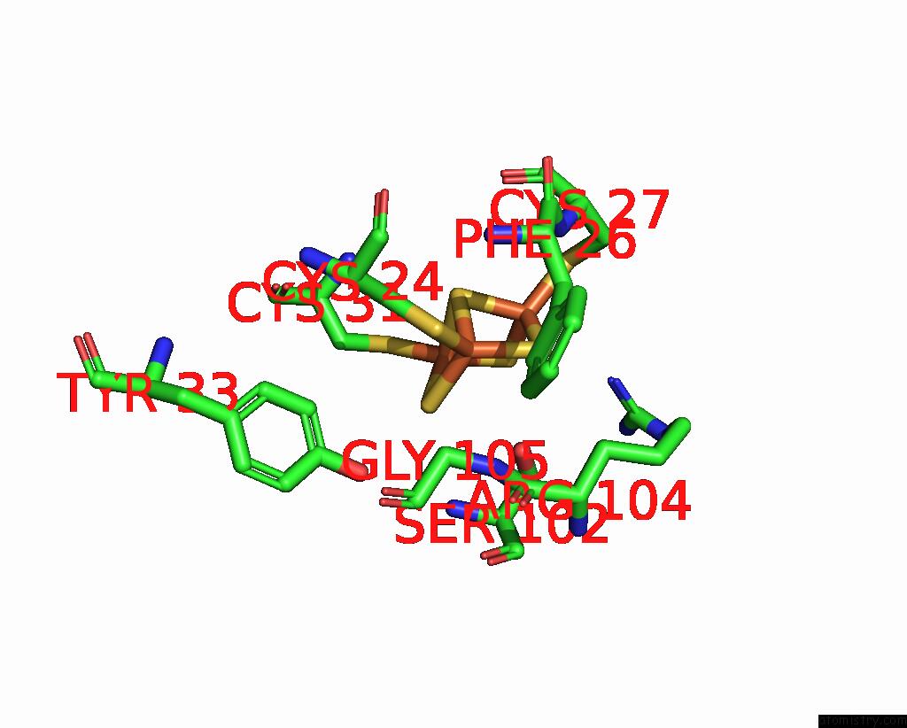
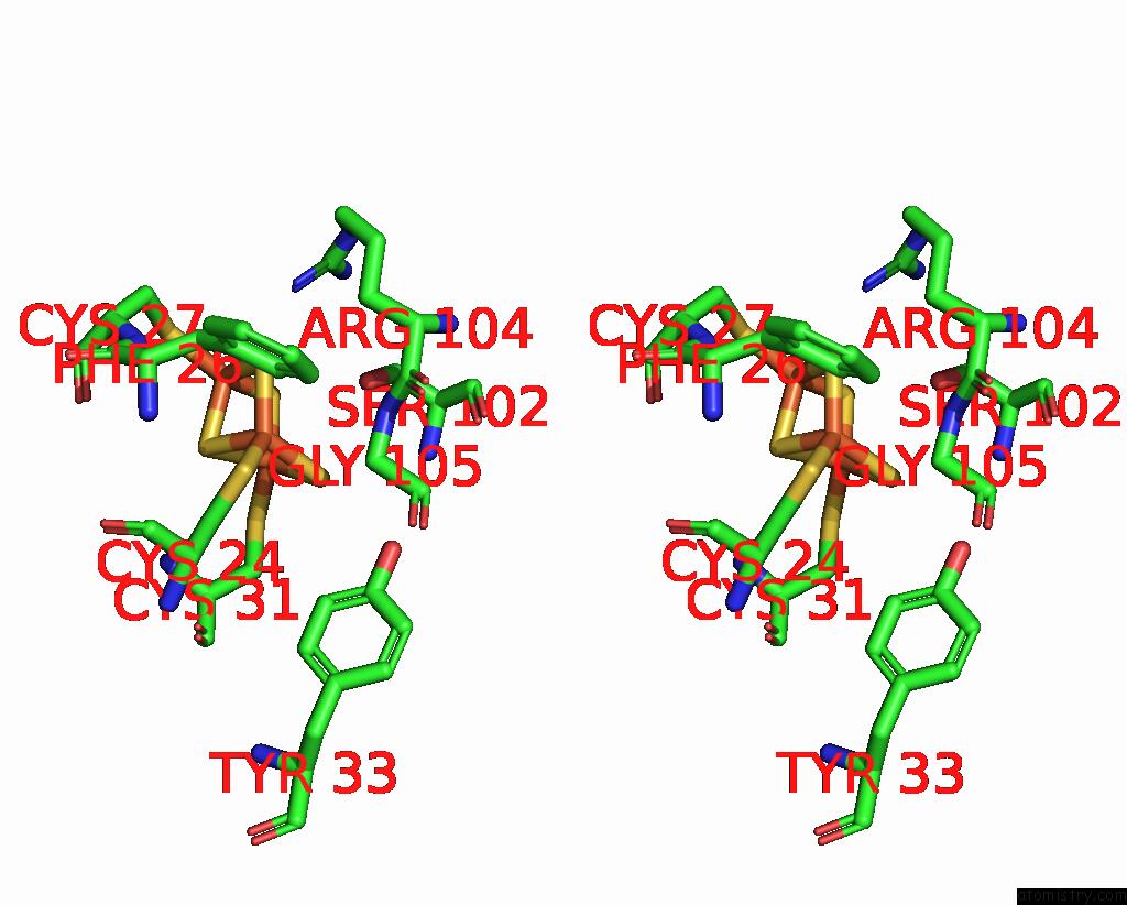
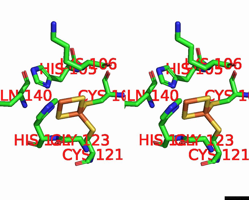
Iron binding site 1 out of 24 in 8ed4
Go back to
Iron binding site 1 out
of 24 in the Structure of the Complex Between the Arsenite Oxidase and Its Native Electron Acceptor Cytochrome C552 From Pseudorhizobium Sp. Str. Nt-26
Mono view

Stereo pair view
Mono view
Stereo pair view
A full contact list of Iron with other atoms in the Fe binding
site number 1 of Structure of the Complex Between the Arsenite Oxidase and Its Native Electron Acceptor Cytochrome C552 From Pseudorhizobium Sp. Str. Nt-26 within 5.0Å range:
|
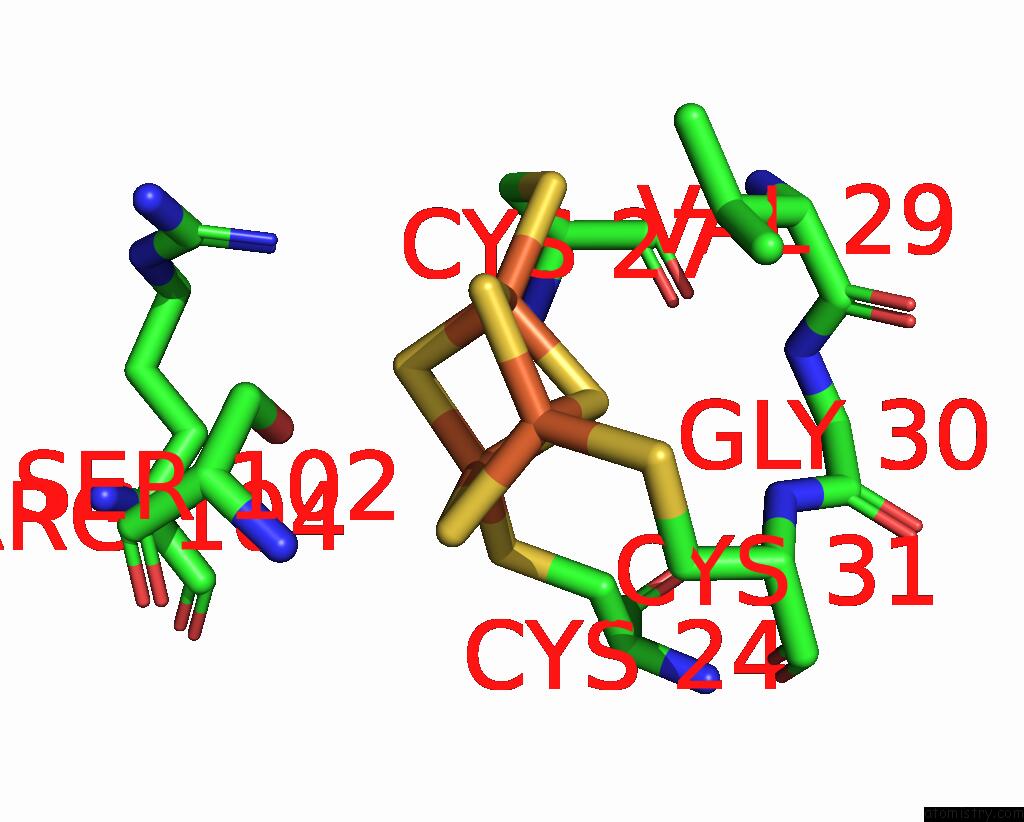
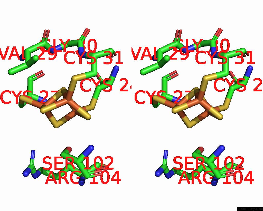
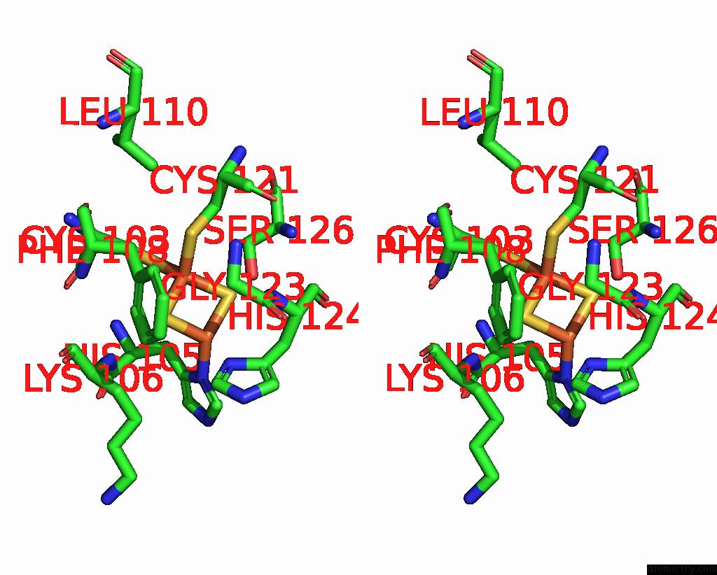
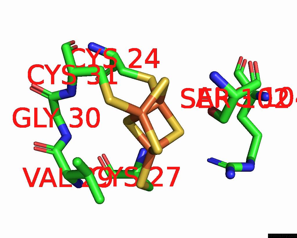
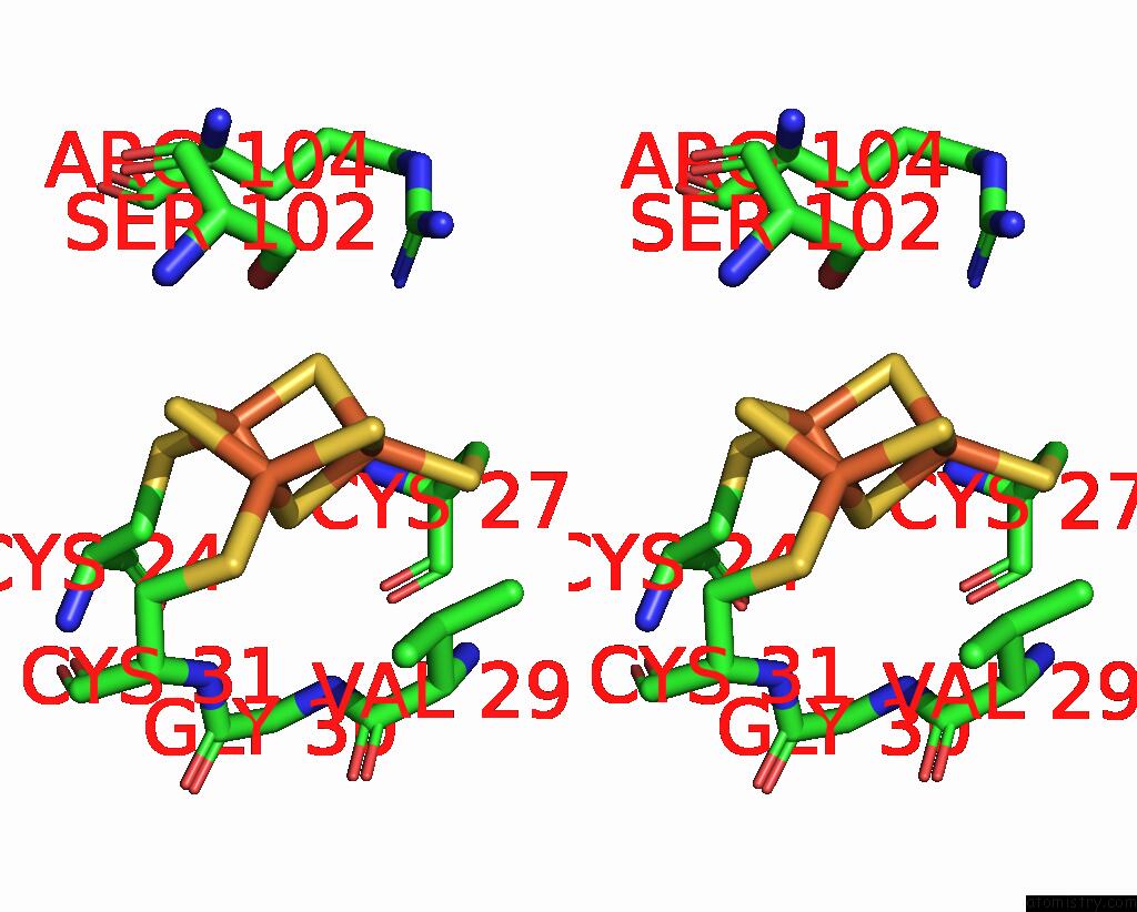
Iron binding site 2 out of 24 in 8ed4
Go back to
Iron binding site 2 out
of 24 in the Structure of the Complex Between the Arsenite Oxidase and Its Native Electron Acceptor Cytochrome C552 From Pseudorhizobium Sp. Str. Nt-26

Mono view
Stereo pair view
Mono view
Stereo pair view
A full contact list of Iron with other atoms in the Fe binding
site number 2 of Structure of the Complex Between the Arsenite Oxidase and Its Native Electron Acceptor Cytochrome C552 From Pseudorhizobium Sp. Str. Nt-26 within 5.0Å range:
|
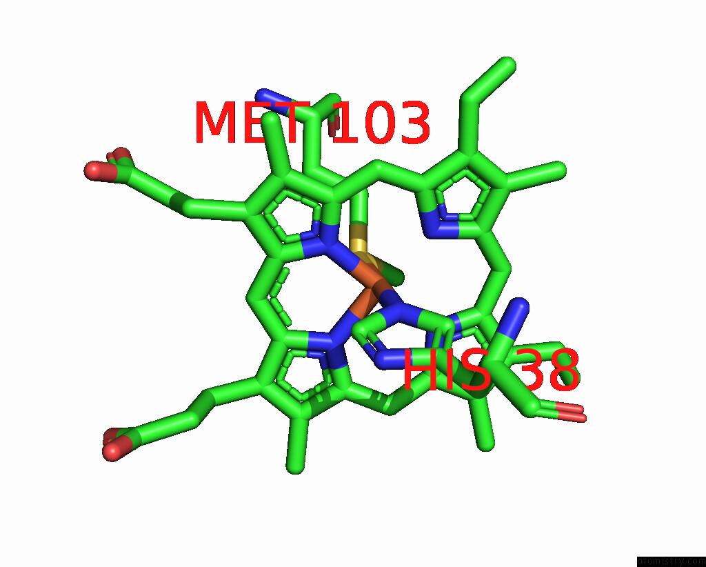
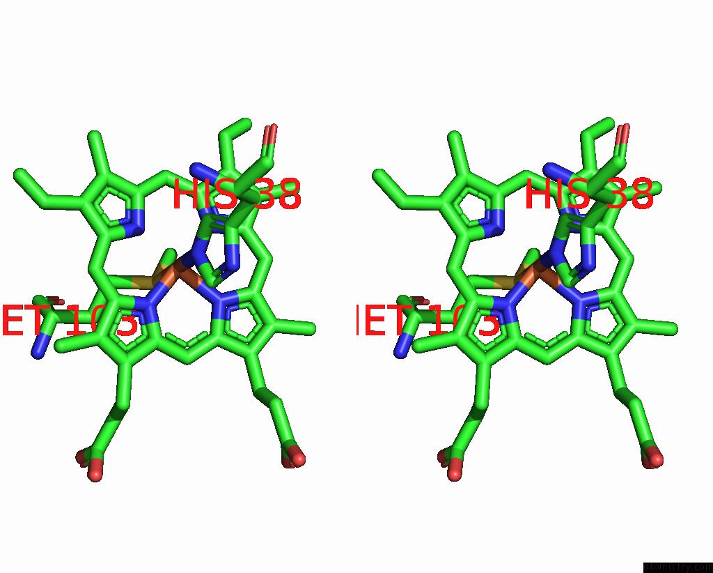
Iron binding site 3 out of 24 in 8ed4
Go back to
Iron binding site 3 out
of 24 in the Structure of the Complex Between the Arsenite Oxidase and Its Native Electron Acceptor Cytochrome C552 From Pseudorhizobium Sp. Str. Nt-26
Mono view
Stereo pair view
Mono view
Stereo pair view
A full contact list of Iron with other atoms in the Fe binding
site number 3 of Structure of the Complex Between the Arsenite Oxidase and Its Native Electron Acceptor Cytochrome C552 From Pseudorhizobium Sp. Str. Nt-26 within 5.0Å range:
|
Iron binding site 4 out of 24 in 8ed4
Go back to
Iron binding site 4 out
of 24 in the Structure of the Complex Between the Arsenite Oxidase and Its Native Electron Acceptor Cytochrome C552 From Pseudorhizobium Sp. Str. Nt-26

Mono view
Stereo pair view
Mono view
Stereo pair view
A full contact list of Iron with other atoms in the Fe binding
site number 4 of Structure of the Complex Between the Arsenite Oxidase and Its Native Electron Acceptor Cytochrome C552 From Pseudorhizobium Sp. Str. Nt-26 within 5.0Å range:
|
Iron binding site 5 out of 24 in 8ed4
Go back to
Iron binding site 5 out
of 24 in the Structure of the Complex Between the Arsenite Oxidase and Its Native Electron Acceptor Cytochrome C552 From Pseudorhizobium Sp. Str. Nt-26

Mono view
Stereo pair view
Mono view
Stereo pair view
A full contact list of Iron with other atoms in the Fe binding
site number 5 of Structure of the Complex Between the Arsenite Oxidase and Its Native Electron Acceptor Cytochrome C552 From Pseudorhizobium Sp. Str. Nt-26 within 5.0Å range:
|
Iron binding site 6 out of 24 in 8ed4
Go back to
Iron binding site 6 out
of 24 in the Structure of the Complex Between the Arsenite Oxidase and Its Native Electron Acceptor Cytochrome C552 From Pseudorhizobium Sp. Str. Nt-26
Mono view
Stereo pair view
Mono view
Stereo pair view
A full contact list of Iron with other atoms in the Fe binding
site number 6 of Structure of the Complex Between the Arsenite Oxidase and Its Native Electron Acceptor Cytochrome C552 From Pseudorhizobium Sp. Str. Nt-26 within 5.0Å range:
|
Iron binding site 7 out of 24 in 8ed4
Go back to
Iron binding site 7 out
of 24 in the Structure of the Complex Between the Arsenite Oxidase and Its Native Electron Acceptor Cytochrome C552 From Pseudorhizobium Sp. Str. Nt-26
Mono view
Stereo pair view
Mono view
Stereo pair view
A full contact list of Iron with other atoms in the Fe binding
site number 7 of Structure of the Complex Between the Arsenite Oxidase and Its Native Electron Acceptor Cytochrome C552 From Pseudorhizobium Sp. Str. Nt-26 within 5.0Å range:
|
Iron binding site 8 out of 24 in 8ed4
Go back to
Iron binding site 8 out
of 24 in the Structure of the Complex Between the Arsenite Oxidase and Its Native Electron Acceptor Cytochrome C552 From Pseudorhizobium Sp. Str. Nt-26

Mono view

Stereo pair view
Mono view
Stereo pair view
A full contact list of Iron with other atoms in the Fe binding
site number 8 of Structure of the Complex Between the Arsenite Oxidase and Its Native Electron Acceptor Cytochrome C552 From Pseudorhizobium Sp. Str. Nt-26 within 5.0Å range:
|
Iron binding site 9 out of 24 in 8ed4
Go back to
Iron binding site 9 out
of 24 in the Structure of the Complex Between the Arsenite Oxidase and Its Native Electron Acceptor Cytochrome C552 From Pseudorhizobium Sp. Str. Nt-26
Mono view
Stereo pair view
Mono view
Stereo pair view
A full contact list of Iron with other atoms in the Fe binding
site number 9 of Structure of the Complex Between the Arsenite Oxidase and Its Native Electron Acceptor Cytochrome C552 From Pseudorhizobium Sp. Str. Nt-26 within 5.0Å range:
|
Iron binding site 10 out of 24 in 8ed4
Go back to
Iron binding site 10 out
of 24 in the Structure of the Complex Between the Arsenite Oxidase and Its Native Electron Acceptor Cytochrome C552 From Pseudorhizobium Sp. Str. Nt-26

Mono view
Stereo pair view
Mono view
Stereo pair view
A full contact list of Iron with other atoms in the Fe binding
site number 10 of Structure of the Complex Between the Arsenite Oxidase and Its Native Electron Acceptor Cytochrome C552 From Pseudorhizobium Sp. Str. Nt-26 within 5.0Å range:
|
Reference:
N.Poddar,
J.M.Santini,
M.J.Maher.
The Structure of the Complex Between the Arsenite Oxidase From Pseudorhizobium Banfieldiae Sp. Strain Nt-26 and Its Native Electron Acceptor Cytochrome C 552. Acta Crystallogr D Struct V. 79 345 2023BIOL.
ISSN: ISSN 2059-7983
PubMed: 36995233
DOI: 10.1107/S2059798323002103
Page generated: Sat Aug 10 01:16:39 2024
ISSN: ISSN 2059-7983
PubMed: 36995233
DOI: 10.1107/S2059798323002103
Last articles
Zn in 9MJ5Zn in 9HNW
Zn in 9G0L
Zn in 9FNE
Zn in 9DZN
Zn in 9E0I
Zn in 9D32
Zn in 9DAK
Zn in 8ZXC
Zn in 8ZUF