Iron »
PDB 8gbt-8hbe »
8gdi »
Iron in PDB 8gdi: X-Ray Crystal Structure of CYP124A1 From Mycobacterium Marinum in Complex with 7-Ketocholesterol
Protein crystallography data
The structure of X-Ray Crystal Structure of CYP124A1 From Mycobacterium Marinum in Complex with 7-Ketocholesterol, PDB code: 8gdi
was solved by
A.Ghith,
J.B.Bruning,
S.G.Bell,
with X-Ray Crystallography technique. A brief refinement statistics is given in the table below:
| Resolution Low / High (Å) | 37.50 / 1.81 |
| Space group | P 21 21 21 |
| Cell size a, b, c (Å), α, β, γ (°) | 67.548, 74.991, 95.078, 90, 90, 90 |
| R / Rfree (%) | 17.5 / 20.7 |
Iron Binding Sites:
The binding sites of Iron atom in the X-Ray Crystal Structure of CYP124A1 From Mycobacterium Marinum in Complex with 7-Ketocholesterol
(pdb code 8gdi). This binding sites where shown within
5.0 Angstroms radius around Iron atom.
In total only one binding site of Iron was determined in the X-Ray Crystal Structure of CYP124A1 From Mycobacterium Marinum in Complex with 7-Ketocholesterol, PDB code: 8gdi:
In total only one binding site of Iron was determined in the X-Ray Crystal Structure of CYP124A1 From Mycobacterium Marinum in Complex with 7-Ketocholesterol, PDB code: 8gdi:
Iron binding site 1 out of 1 in 8gdi
Go back to
Iron binding site 1 out
of 1 in the X-Ray Crystal Structure of CYP124A1 From Mycobacterium Marinum in Complex with 7-Ketocholesterol

Mono view
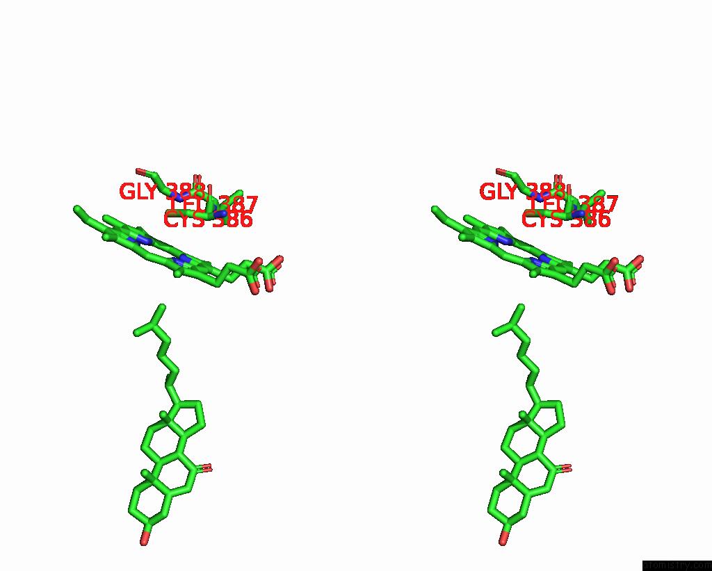
Stereo pair view
Mono view
Stereo pair view
A full contact list of Iron with other atoms in the Fe binding
site number 1 of X-Ray Crystal Structure of CYP124A1 From Mycobacterium Marinum in Complex with 7-Ketocholesterol within 5.0Å range:
|
Reference:
A.Ghith,
J.B.Bruning,
S.G.Bell.
The Oxidation of Cholesterol Derivatives By the CYP124 and CYP142 Enzymes From Mycobacterium Marinum. J.Steroid Biochem.Mol.Biol. V. 231 06317 2023.
ISSN: ISSN 0960-0760
PubMed: 37141947
DOI: 10.1016/J.JSBMB.2023.106317
Page generated: Sat Aug 10 04:51:07 2024
ISSN: ISSN 0960-0760
PubMed: 37141947
DOI: 10.1016/J.JSBMB.2023.106317
Last articles
Zn in 9J0NZn in 9J0O
Zn in 9J0P
Zn in 9FJX
Zn in 9EKB
Zn in 9C0F
Zn in 9CAH
Zn in 9CH0
Zn in 9CH3
Zn in 9CH1