Iron »
PDB 8gbt-8hbe »
8grj »
Iron in PDB 8grj: Crystal Structure of Gamma-Alpha Subunit Complex From Burkholderia Cepacia Fad Glucose Dehydrogenase in Complex with Gluconolactone
Protein crystallography data
The structure of Crystal Structure of Gamma-Alpha Subunit Complex From Burkholderia Cepacia Fad Glucose Dehydrogenase in Complex with Gluconolactone, PDB code: 8grj
was solved by
H.Yoshida,
K.Kojima,
W.Tsugawa,
J.Okuda-Shimazaki,
J.A.Kerrigan,
K.Sode,
with X-Ray Crystallography technique. A brief refinement statistics is given in the table below:
| Resolution Low / High (Å) | 50.04 / 2.95 |
| Space group | P 65 2 2 |
| Cell size a, b, c (Å), α, β, γ (°) | 111.235, 111.235, 527.106, 90, 90, 120 |
| R / Rfree (%) | 19.8 / 27 |
Iron Binding Sites:
The binding sites of Iron atom in the Crystal Structure of Gamma-Alpha Subunit Complex From Burkholderia Cepacia Fad Glucose Dehydrogenase in Complex with Gluconolactone
(pdb code 8grj). This binding sites where shown within
5.0 Angstroms radius around Iron atom.
In total 6 binding sites of Iron where determined in the Crystal Structure of Gamma-Alpha Subunit Complex From Burkholderia Cepacia Fad Glucose Dehydrogenase in Complex with Gluconolactone, PDB code: 8grj:
Jump to Iron binding site number: 1; 2; 3; 4; 5; 6;
In total 6 binding sites of Iron where determined in the Crystal Structure of Gamma-Alpha Subunit Complex From Burkholderia Cepacia Fad Glucose Dehydrogenase in Complex with Gluconolactone, PDB code: 8grj:
Jump to Iron binding site number: 1; 2; 3; 4; 5; 6;
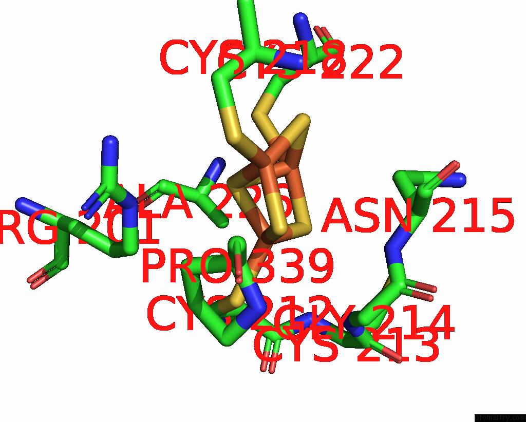
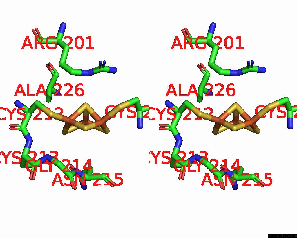
Iron binding site 1 out of 6 in 8grj
Go back to
Iron binding site 1 out
of 6 in the Crystal Structure of Gamma-Alpha Subunit Complex From Burkholderia Cepacia Fad Glucose Dehydrogenase in Complex with Gluconolactone
Mono view

Stereo pair view
Mono view
Stereo pair view
A full contact list of Iron with other atoms in the Fe binding
site number 1 of Crystal Structure of Gamma-Alpha Subunit Complex From Burkholderia Cepacia Fad Glucose Dehydrogenase in Complex with Gluconolactone within 5.0Å range:
|
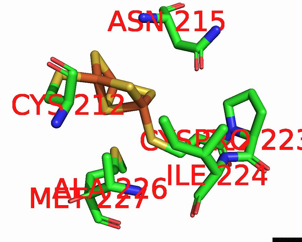
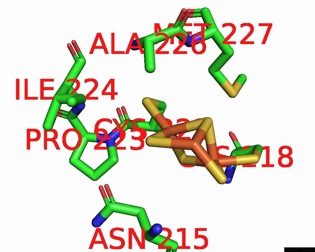
Iron binding site 2 out of 6 in 8grj
Go back to
Iron binding site 2 out
of 6 in the Crystal Structure of Gamma-Alpha Subunit Complex From Burkholderia Cepacia Fad Glucose Dehydrogenase in Complex with Gluconolactone
Mono view

Stereo pair view
Mono view
Stereo pair view
A full contact list of Iron with other atoms in the Fe binding
site number 2 of Crystal Structure of Gamma-Alpha Subunit Complex From Burkholderia Cepacia Fad Glucose Dehydrogenase in Complex with Gluconolactone within 5.0Å range:
|
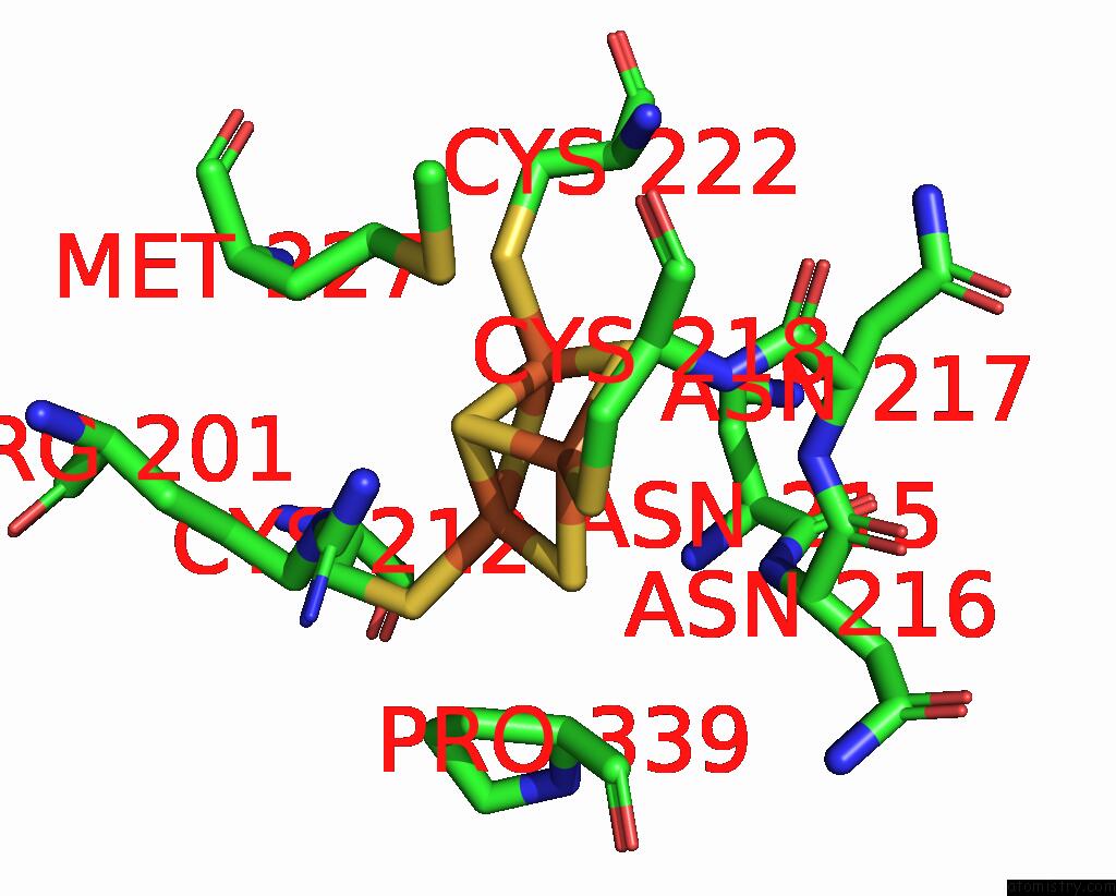
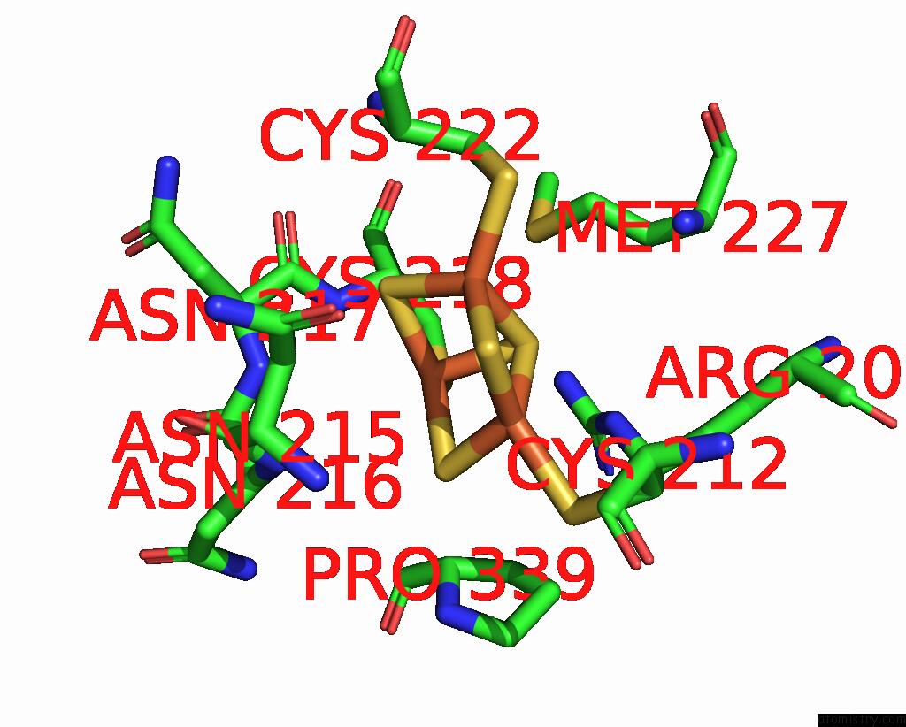
Iron binding site 3 out of 6 in 8grj
Go back to
Iron binding site 3 out
of 6 in the Crystal Structure of Gamma-Alpha Subunit Complex From Burkholderia Cepacia Fad Glucose Dehydrogenase in Complex with Gluconolactone
Mono view

Stereo pair view
Mono view
Stereo pair view
A full contact list of Iron with other atoms in the Fe binding
site number 3 of Crystal Structure of Gamma-Alpha Subunit Complex From Burkholderia Cepacia Fad Glucose Dehydrogenase in Complex with Gluconolactone within 5.0Å range:
|
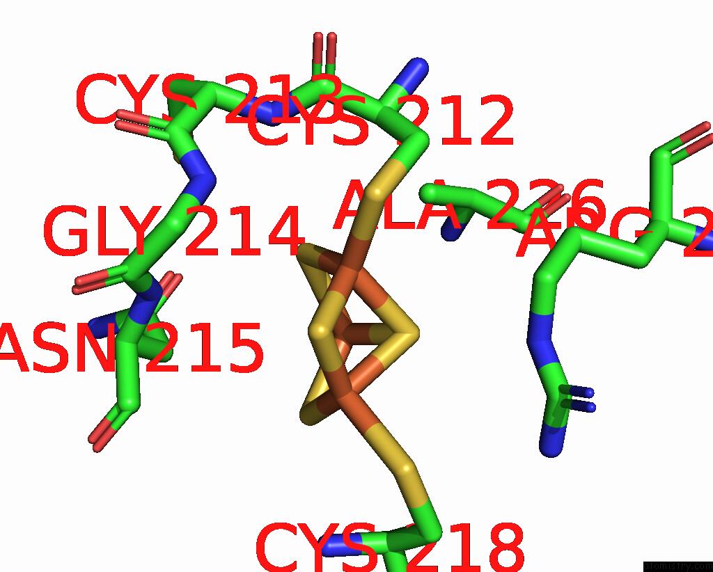
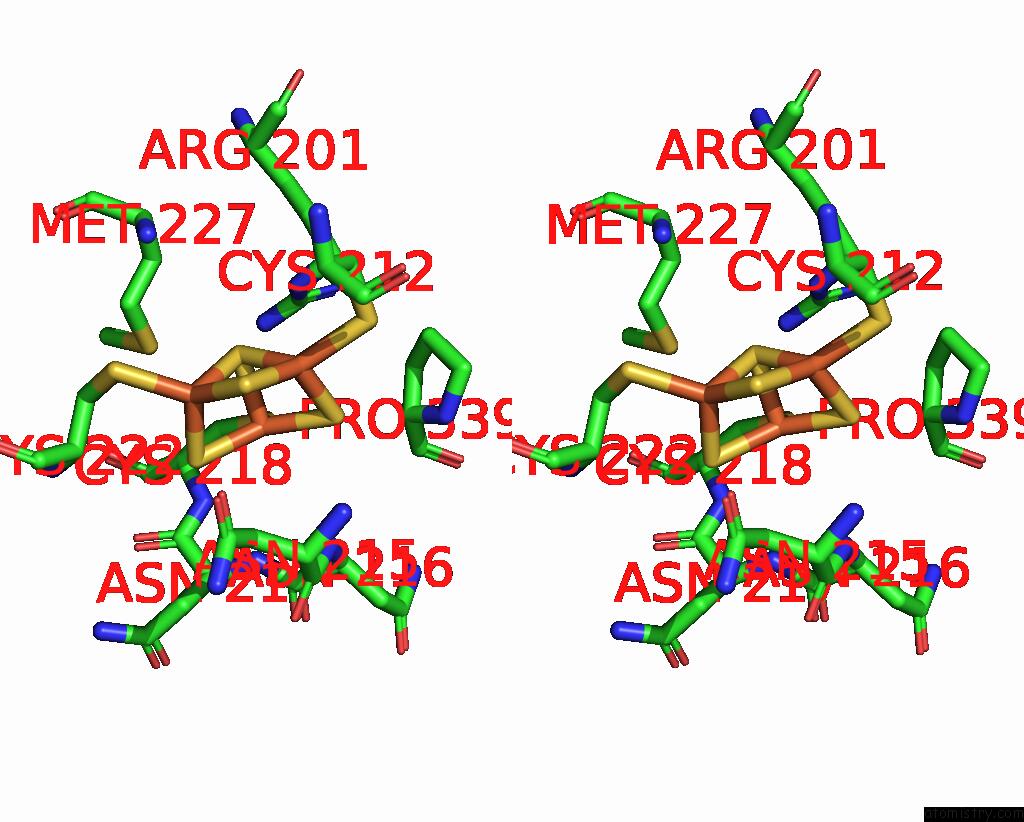
Iron binding site 4 out of 6 in 8grj
Go back to
Iron binding site 4 out
of 6 in the Crystal Structure of Gamma-Alpha Subunit Complex From Burkholderia Cepacia Fad Glucose Dehydrogenase in Complex with Gluconolactone
Mono view
Stereo pair view
Mono view
Stereo pair view
A full contact list of Iron with other atoms in the Fe binding
site number 4 of Crystal Structure of Gamma-Alpha Subunit Complex From Burkholderia Cepacia Fad Glucose Dehydrogenase in Complex with Gluconolactone within 5.0Å range:
|
Iron binding site 5 out of 6 in 8grj
Go back to
Iron binding site 5 out
of 6 in the Crystal Structure of Gamma-Alpha Subunit Complex From Burkholderia Cepacia Fad Glucose Dehydrogenase in Complex with Gluconolactone
Mono view

Stereo pair view
Mono view
Stereo pair view
A full contact list of Iron with other atoms in the Fe binding
site number 5 of Crystal Structure of Gamma-Alpha Subunit Complex From Burkholderia Cepacia Fad Glucose Dehydrogenase in Complex with Gluconolactone within 5.0Å range:
|
Iron binding site 6 out of 6 in 8grj
Go back to
Iron binding site 6 out
of 6 in the Crystal Structure of Gamma-Alpha Subunit Complex From Burkholderia Cepacia Fad Glucose Dehydrogenase in Complex with Gluconolactone
Mono view
Stereo pair view
Mono view
Stereo pair view
A full contact list of Iron with other atoms in the Fe binding
site number 6 of Crystal Structure of Gamma-Alpha Subunit Complex From Burkholderia Cepacia Fad Glucose Dehydrogenase in Complex with Gluconolactone within 5.0Å range:
|
Reference:
H.Yoshida,
K.Kojima,
W.Tsugawa,
J.Okuda-Shimazaki,
J.A.Kerrigan,
K.Sode.
Crystal Structure of Gamma-Alpha Subunit Complex From Burkholderia Cepacia Fad Glucose Dehydrogenase in Complex with Gluconolactone To Be Published.
Page generated: Sat Aug 10 04:57:54 2024
Last articles
Zn in 9J0NZn in 9J0O
Zn in 9J0P
Zn in 9FJX
Zn in 9EKB
Zn in 9C0F
Zn in 9CAH
Zn in 9CH0
Zn in 9CH3
Zn in 9CH1