Iron »
PDB 8gbt-8hbe »
8gtl »
Iron in PDB 8gtl: Crystal Structure of Cytochrome P450 (CYP101D5)
Protein crystallography data
The structure of Crystal Structure of Cytochrome P450 (CYP101D5), PDB code: 8gtl
was solved by
H.Do,
J.H.Lee,
with X-Ray Crystallography technique. A brief refinement statistics is given in the table below:
| Resolution Low / High (Å) | 43.79 / 3.20 |
| Space group | P 21 21 21 |
| Cell size a, b, c (Å), α, β, γ (°) | 68.524, 109.573, 113.878, 90, 90, 90 |
| R / Rfree (%) | 26.2 / 31.9 |
Iron Binding Sites:
The binding sites of Iron atom in the Crystal Structure of Cytochrome P450 (CYP101D5)
(pdb code 8gtl). This binding sites where shown within
5.0 Angstroms radius around Iron atom.
In total 2 binding sites of Iron where determined in the Crystal Structure of Cytochrome P450 (CYP101D5), PDB code: 8gtl:
Jump to Iron binding site number: 1; 2;
In total 2 binding sites of Iron where determined in the Crystal Structure of Cytochrome P450 (CYP101D5), PDB code: 8gtl:
Jump to Iron binding site number: 1; 2;
Iron binding site 1 out of 2 in 8gtl
Go back to
Iron binding site 1 out
of 2 in the Crystal Structure of Cytochrome P450 (CYP101D5)
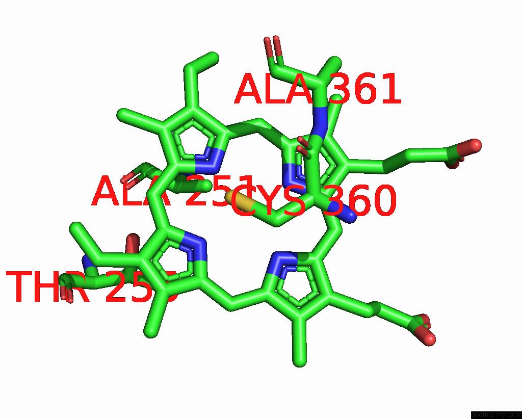
Mono view
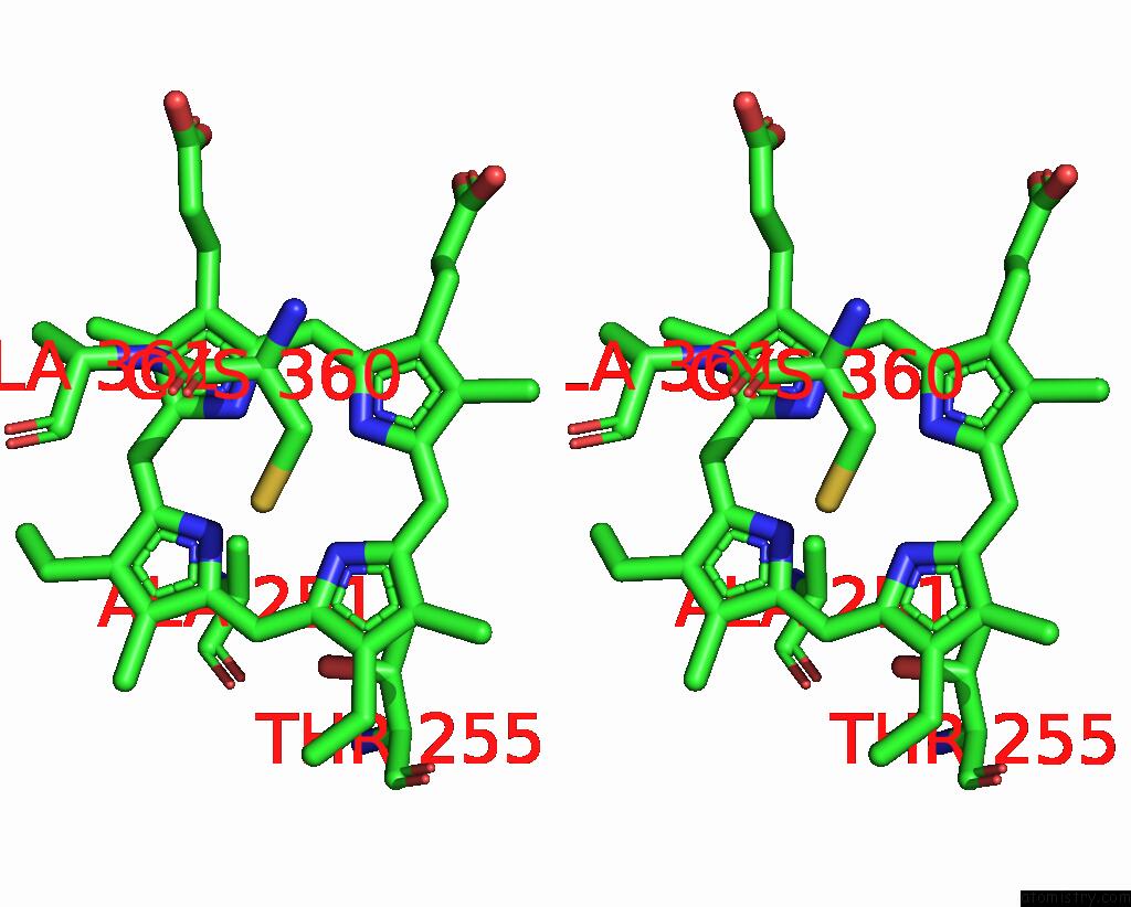
Stereo pair view
Mono view
Stereo pair view
A full contact list of Iron with other atoms in the Fe binding
site number 1 of Crystal Structure of Cytochrome P450 (CYP101D5) within 5.0Å range:
|
Iron binding site 2 out of 2 in 8gtl
Go back to
Iron binding site 2 out
of 2 in the Crystal Structure of Cytochrome P450 (CYP101D5)
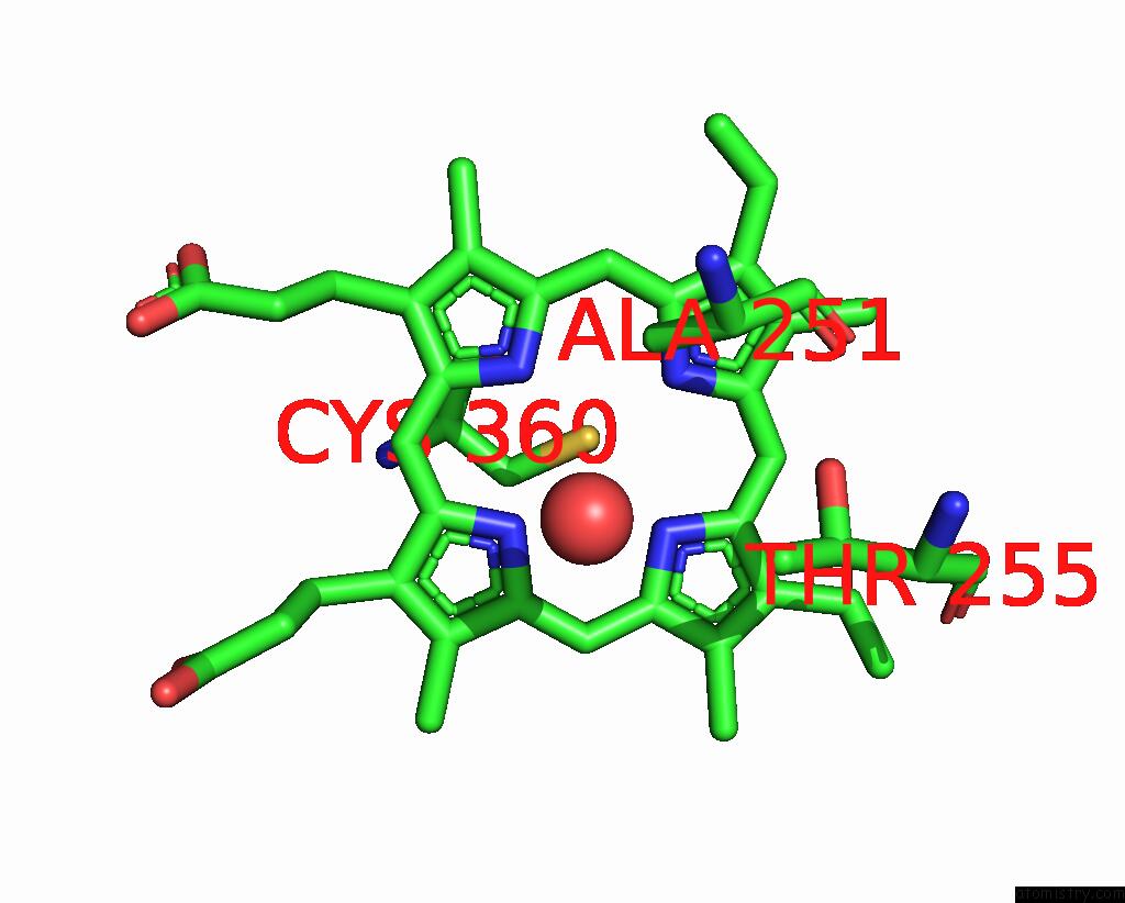
Mono view
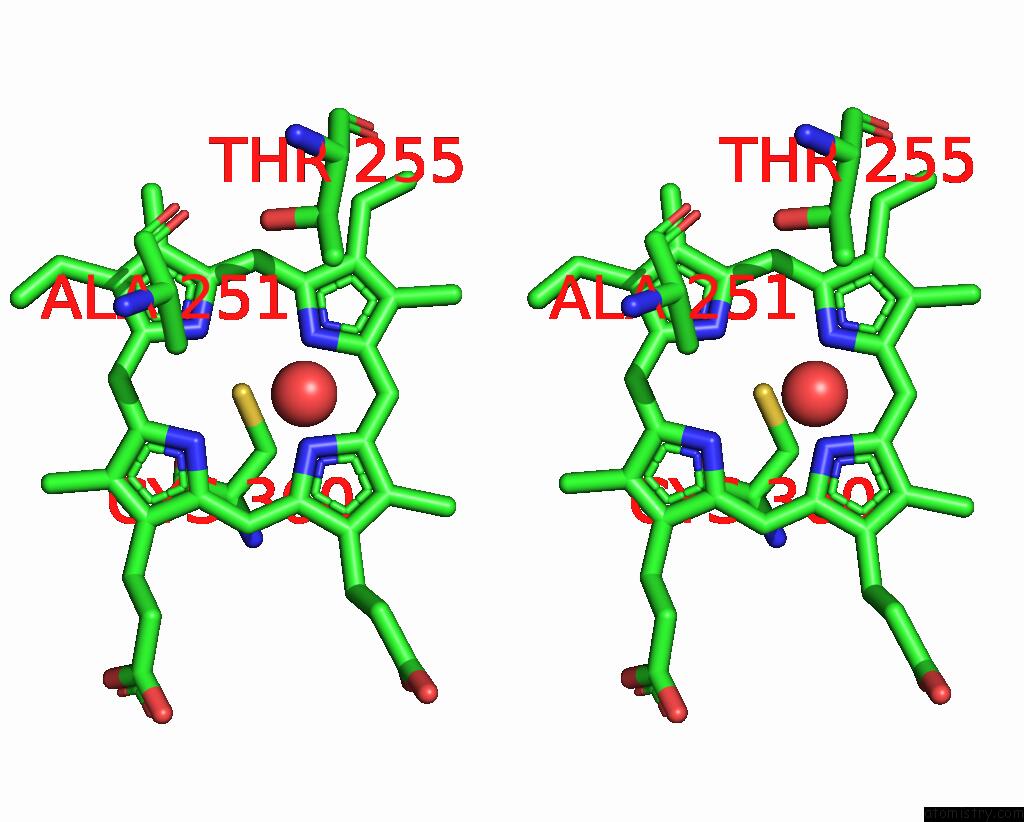
Stereo pair view
Mono view
Stereo pair view
A full contact list of Iron with other atoms in the Fe binding
site number 2 of Crystal Structure of Cytochrome P450 (CYP101D5) within 5.0Å range:
|
Reference:
P.Subedi,
H.Do,
J.H.Lee,
T.J.Oh.
Crystal Structure and Biochemical Analysis of A Cytochrome P450 CYP101D5 From Sphingomonas Echinoides. Int J Mol Sci V. 23 2022.
ISSN: ESSN 1422-0067
PubMed: 36362105
DOI: 10.3390/IJMS232113317
Page generated: Sat Aug 10 05:00:31 2024
ISSN: ESSN 1422-0067
PubMed: 36362105
DOI: 10.3390/IJMS232113317
Last articles
Zn in 9J0NZn in 9J0O
Zn in 9J0P
Zn in 9FJX
Zn in 9EKB
Zn in 9C0F
Zn in 9CAH
Zn in 9CH0
Zn in 9CH3
Zn in 9CH1