Iron »
PDB 8gbt-8hbe »
8h7y »
Iron in PDB 8h7y: Trans-3/4-Proline-Hydroxylase H11 with Akg and L-Proline
Protein crystallography data
The structure of Trans-3/4-Proline-Hydroxylase H11 with Akg and L-Proline, PDB code: 8h7y
was solved by
W.M.Gong,
X.Y.Hu,
with X-Ray Crystallography technique. A brief refinement statistics is given in the table below:
| Resolution Low / High (Å) | 39.79 / 2.14 |
| Space group | P 41 21 2 |
| Cell size a, b, c (Å), α, β, γ (°) | 106.437, 106.437, 145.016, 90, 90, 90 |
| R / Rfree (%) | 15.2 / 21.6 |
Iron Binding Sites:
The binding sites of Iron atom in the Trans-3/4-Proline-Hydroxylase H11 with Akg and L-Proline
(pdb code 8h7y). This binding sites where shown within
5.0 Angstroms radius around Iron atom.
In total 2 binding sites of Iron where determined in the Trans-3/4-Proline-Hydroxylase H11 with Akg and L-Proline, PDB code: 8h7y:
Jump to Iron binding site number: 1; 2;
In total 2 binding sites of Iron where determined in the Trans-3/4-Proline-Hydroxylase H11 with Akg and L-Proline, PDB code: 8h7y:
Jump to Iron binding site number: 1; 2;
Iron binding site 1 out of 2 in 8h7y
Go back to
Iron binding site 1 out
of 2 in the Trans-3/4-Proline-Hydroxylase H11 with Akg and L-Proline
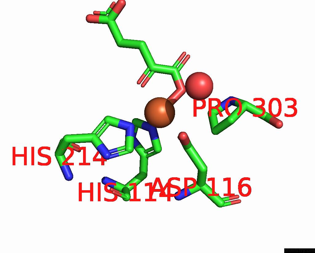
Mono view
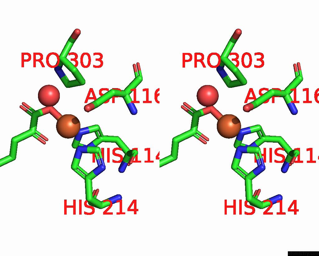
Stereo pair view
Mono view
Stereo pair view
A full contact list of Iron with other atoms in the Fe binding
site number 1 of Trans-3/4-Proline-Hydroxylase H11 with Akg and L-Proline within 5.0Å range:
|
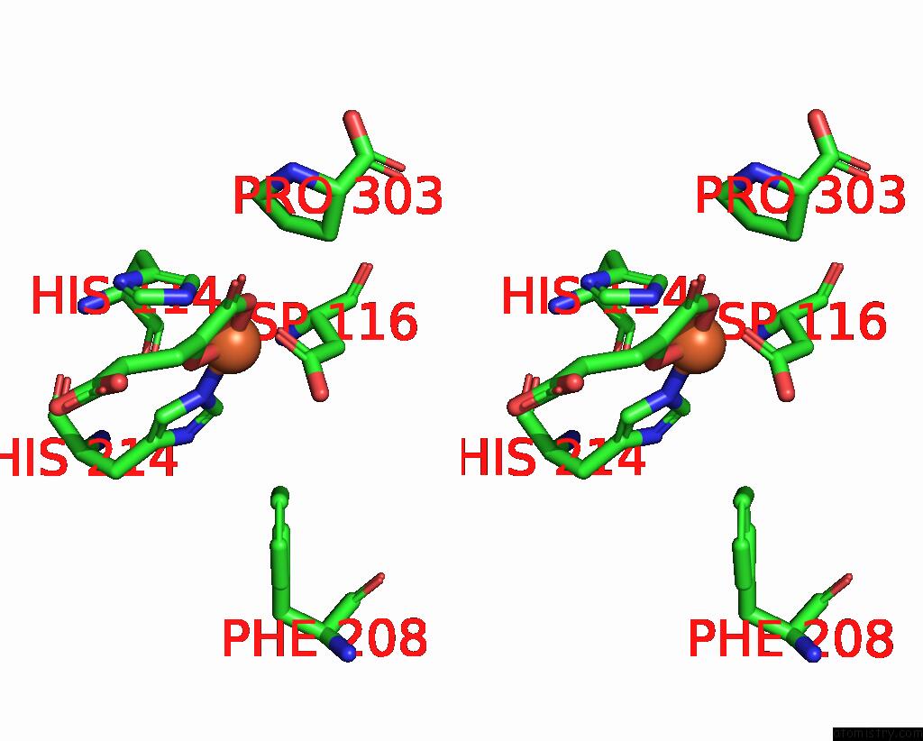
Iron binding site 2 out of 2 in 8h7y
Go back to
Iron binding site 2 out
of 2 in the Trans-3/4-Proline-Hydroxylase H11 with Akg and L-Proline

Mono view
Stereo pair view
Mono view
Stereo pair view
A full contact list of Iron with other atoms in the Fe binding
site number 2 of Trans-3/4-Proline-Hydroxylase H11 with Akg and L-Proline within 5.0Å range:
|
Reference:
X.Hu,
X.Huang,
J.Liu,
P.Zheng,
W.Gong,
L.Yang.
Structures of L-Proline Trans-Hydroxylase Reveal the Catalytic Specificity and Provide Deeper Insight Into Akg-Dependent Hydroxylation. Acta Crystallogr D Struct V. 79 318 2023BIOL.
ISSN: ISSN 2059-7983
PubMed: 36974966
DOI: 10.1107/S2059798323001936
Page generated: Sat Aug 10 05:08:54 2024
ISSN: ISSN 2059-7983
PubMed: 36974966
DOI: 10.1107/S2059798323001936
Last articles
Cl in 5RB3Cl in 5RB2
Cl in 5RB0
Cl in 5RB1
Cl in 5RAZ
Cl in 5RAY
Cl in 5RAX
Cl in 5RAW
Cl in 5RAV
Cl in 5RAU