Iron »
PDB 8k6j-8ovd »
8one »
Iron in PDB 8one: Crystal Structure of Full-Length Human Lysyl Hydroxylase LH3 - ASP190SER Mutant - Cocrystal with FE2+, MN2+, Udp-Glucose
Enzymatic activity of Crystal Structure of Full-Length Human Lysyl Hydroxylase LH3 - ASP190SER Mutant - Cocrystal with FE2+, MN2+, Udp-Glucose
All present enzymatic activity of Crystal Structure of Full-Length Human Lysyl Hydroxylase LH3 - ASP190SER Mutant - Cocrystal with FE2+, MN2+, Udp-Glucose:
1.14.11.4; 2.4.1.50; 2.4.1.66;
1.14.11.4; 2.4.1.50; 2.4.1.66;
Protein crystallography data
The structure of Crystal Structure of Full-Length Human Lysyl Hydroxylase LH3 - ASP190SER Mutant - Cocrystal with FE2+, MN2+, Udp-Glucose, PDB code: 8one
was solved by
D.Mattoteia,
M.De Marco,
A.Pinnola,
S.Faravelli,
L.Scietti,
F.Forneris,
with X-Ray Crystallography technique. A brief refinement statistics is given in the table below:
| Resolution Low / High (Å) | 47.44 / 2.30 |
| Space group | C 2 2 21 |
| Cell size a, b, c (Å), α, β, γ (°) | 97.09, 100.181, 223.794, 90, 90, 90 |
| R / Rfree (%) | 20.4 / 22.8 |
Other elements in 8one:
The structure of Crystal Structure of Full-Length Human Lysyl Hydroxylase LH3 - ASP190SER Mutant - Cocrystal with FE2+, MN2+, Udp-Glucose also contains other interesting chemical elements:
| Manganese | (Mn) | 1 atom |
Iron Binding Sites:
The binding sites of Iron atom in the Crystal Structure of Full-Length Human Lysyl Hydroxylase LH3 - ASP190SER Mutant - Cocrystal with FE2+, MN2+, Udp-Glucose
(pdb code 8one). This binding sites where shown within
5.0 Angstroms radius around Iron atom.
In total 2 binding sites of Iron where determined in the Crystal Structure of Full-Length Human Lysyl Hydroxylase LH3 - ASP190SER Mutant - Cocrystal with FE2+, MN2+, Udp-Glucose, PDB code: 8one:
Jump to Iron binding site number: 1; 2;
In total 2 binding sites of Iron where determined in the Crystal Structure of Full-Length Human Lysyl Hydroxylase LH3 - ASP190SER Mutant - Cocrystal with FE2+, MN2+, Udp-Glucose, PDB code: 8one:
Jump to Iron binding site number: 1; 2;
Iron binding site 1 out of 2 in 8one
Go back to
Iron binding site 1 out
of 2 in the Crystal Structure of Full-Length Human Lysyl Hydroxylase LH3 - ASP190SER Mutant - Cocrystal with FE2+, MN2+, Udp-Glucose
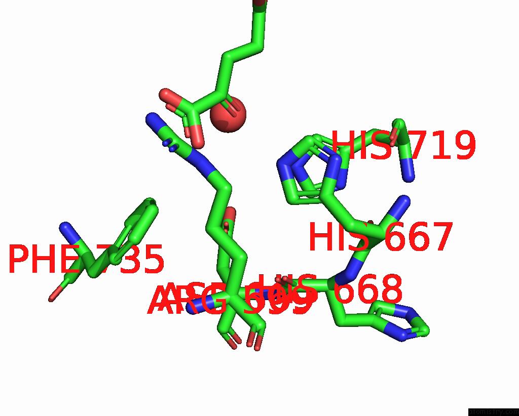
Mono view
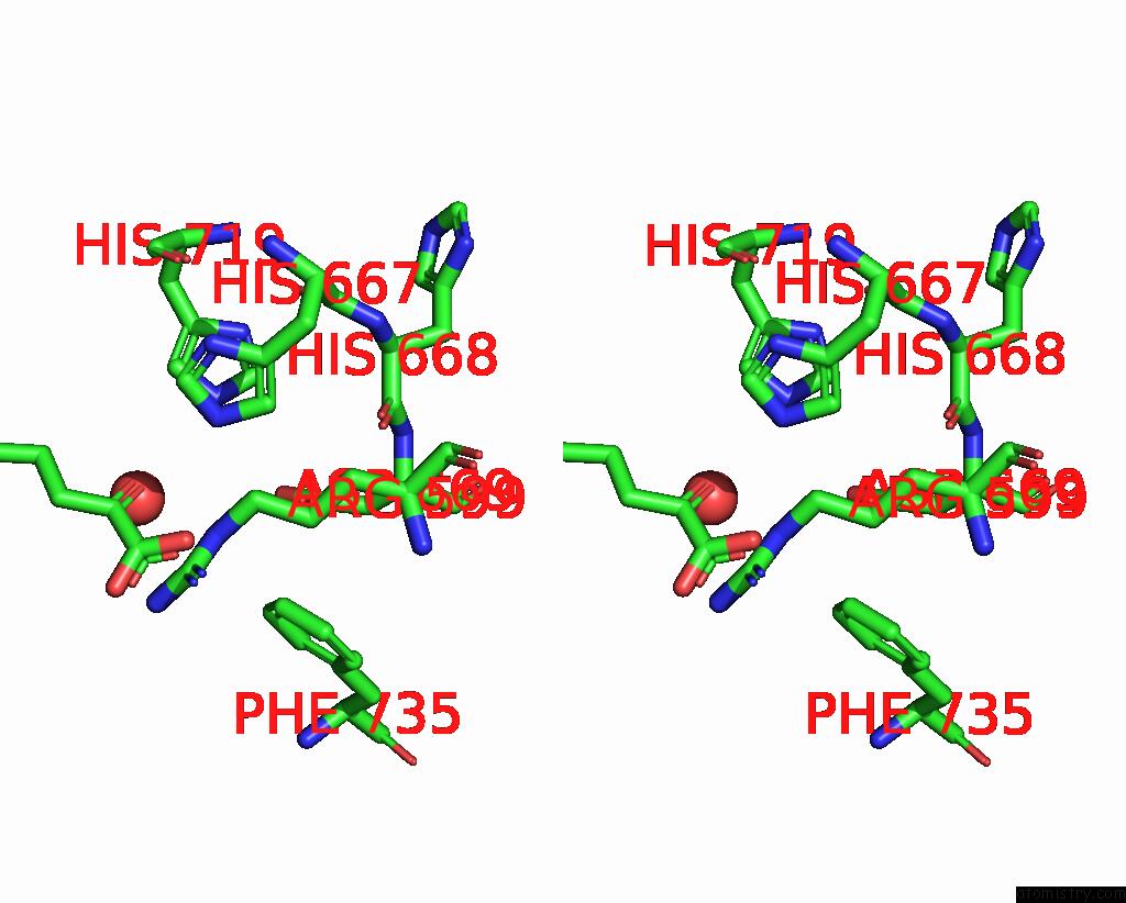
Stereo pair view
Mono view
Stereo pair view
A full contact list of Iron with other atoms in the Fe binding
site number 1 of Crystal Structure of Full-Length Human Lysyl Hydroxylase LH3 - ASP190SER Mutant - Cocrystal with FE2+, MN2+, Udp-Glucose within 5.0Å range:
|
Iron binding site 2 out of 2 in 8one
Go back to
Iron binding site 2 out
of 2 in the Crystal Structure of Full-Length Human Lysyl Hydroxylase LH3 - ASP190SER Mutant - Cocrystal with FE2+, MN2+, Udp-Glucose
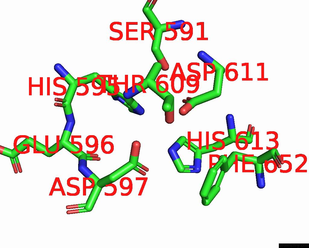
Mono view
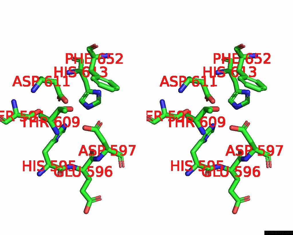
Stereo pair view
Mono view
Stereo pair view
A full contact list of Iron with other atoms in the Fe binding
site number 2 of Crystal Structure of Full-Length Human Lysyl Hydroxylase LH3 - ASP190SER Mutant - Cocrystal with FE2+, MN2+, Udp-Glucose within 5.0Å range:
|
Reference:
D.Mattoteia,
A.Chiapparino,
M.Fumagalli,
M.De Marco,
F.De Giorgi,
L.Negro,
A.Pinnola,
S.Faravelli,
T.Roscioli,
L.Scietti,
F.Forneris.
Identification of Regulatory Molecular 'Hot Spots' For Lh/Plod Collagen Glycosyltransferase Activity Int J Mol Sci 2023.
ISSN: ESSN 1422-0067
DOI: 10.3390/IJMS241311213
Page generated: Sat Aug 10 08:24:29 2024
ISSN: ESSN 1422-0067
DOI: 10.3390/IJMS241311213
Last articles
Zn in 9J0NZn in 9J0O
Zn in 9J0P
Zn in 9FJX
Zn in 9EKB
Zn in 9C0F
Zn in 9CAH
Zn in 9CH0
Zn in 9CH3
Zn in 9CH1