Iron »
PDB 8oz5-8pxq »
8poz »
Iron in PDB 8poz: Crystal Structure of the C120G Variant of the Membrane-Bound [Nife]- Hydrogenase From Cupriavidus Necator in the H2-Reduced State at 1.65 A Resolution.
Enzymatic activity of Crystal Structure of the C120G Variant of the Membrane-Bound [Nife]- Hydrogenase From Cupriavidus Necator in the H2-Reduced State at 1.65 A Resolution.
All present enzymatic activity of Crystal Structure of the C120G Variant of the Membrane-Bound [Nife]- Hydrogenase From Cupriavidus Necator in the H2-Reduced State at 1.65 A Resolution.:
1.12.99.6;
1.12.99.6;
Protein crystallography data
The structure of Crystal Structure of the C120G Variant of the Membrane-Bound [Nife]- Hydrogenase From Cupriavidus Necator in the H2-Reduced State at 1.65 A Resolution., PDB code: 8poz
was solved by
A.Schmidt,
J.Kalms,
P.Scheerer,
with X-Ray Crystallography technique. A brief refinement statistics is given in the table below:
| Resolution Low / High (Å) | 47.92 / 1.65 |
| Space group | P 21 21 21 |
| Cell size a, b, c (Å), α, β, γ (°) | 73.415, 95.751, 120.597, 90, 90, 90 |
| R / Rfree (%) | 14.1 / 17.2 |
Other elements in 8poz:
The structure of Crystal Structure of the C120G Variant of the Membrane-Bound [Nife]- Hydrogenase From Cupriavidus Necator in the H2-Reduced State at 1.65 A Resolution. also contains other interesting chemical elements:
| Magnesium | (Mg) | 1 atom |
| Nickel | (Ni) | 1 atom |
| Chlorine | (Cl) | 4 atoms |
Iron Binding Sites:
Pages:
>>> Page 1 <<< Page 2, Binding sites: 11 - 16;Binding sites:
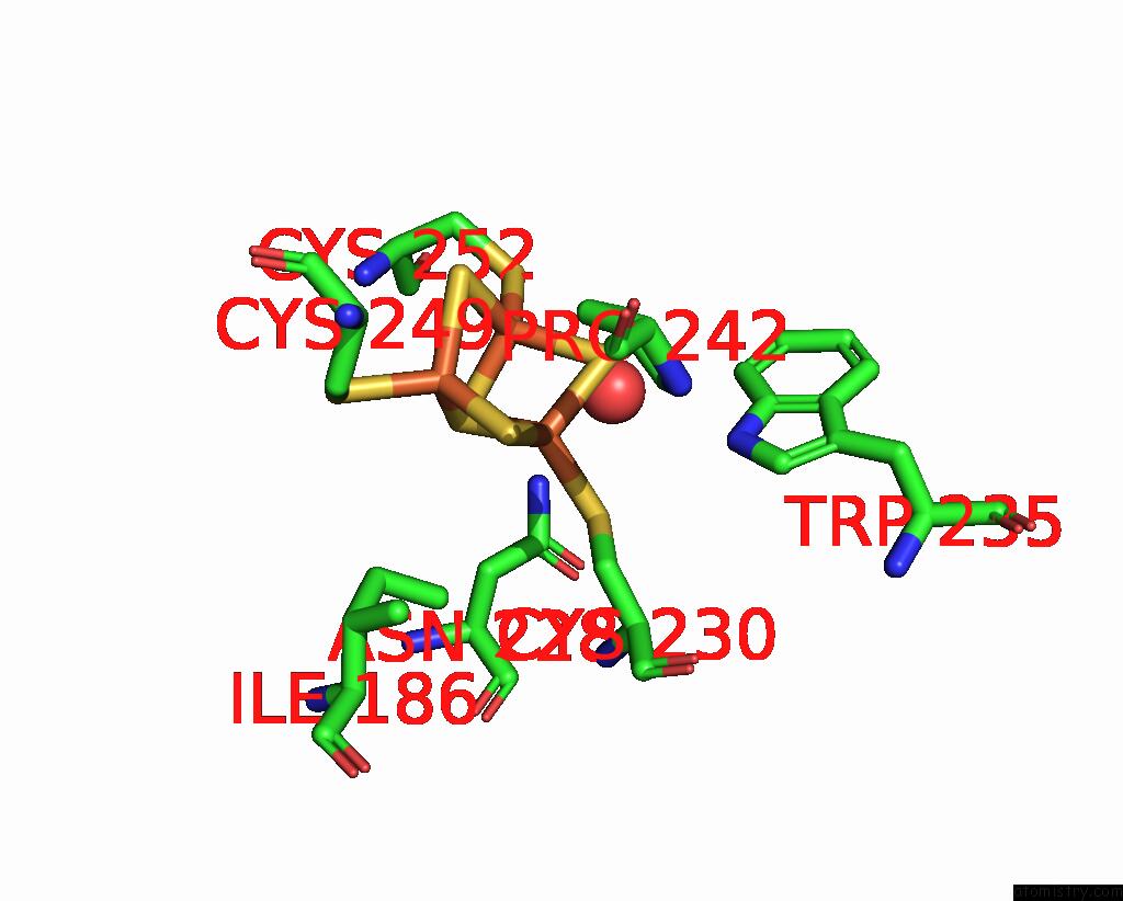
The binding sites of Iron atom in the Crystal Structure of the C120G Variant of the Membrane-Bound [Nife]- Hydrogenase From Cupriavidus Necator in the H2-Reduced State at 1.65 A Resolution. (pdb code 8poz). This binding sites where shown within 5.0 Angstroms radius around Iron atom.In total 16 binding sites of Iron where determined in the Crystal Structure of the C120G Variant of the Membrane-Bound [Nife]- Hydrogenase From Cupriavidus Necator in the H2-Reduced State at 1.65 A Resolution., PDB code: 8poz:
Jump to Iron binding site number: 1; 2; 3; 4; 5; 6; 7; 8; 9; 10;
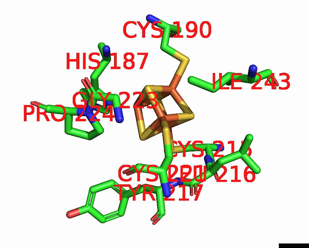
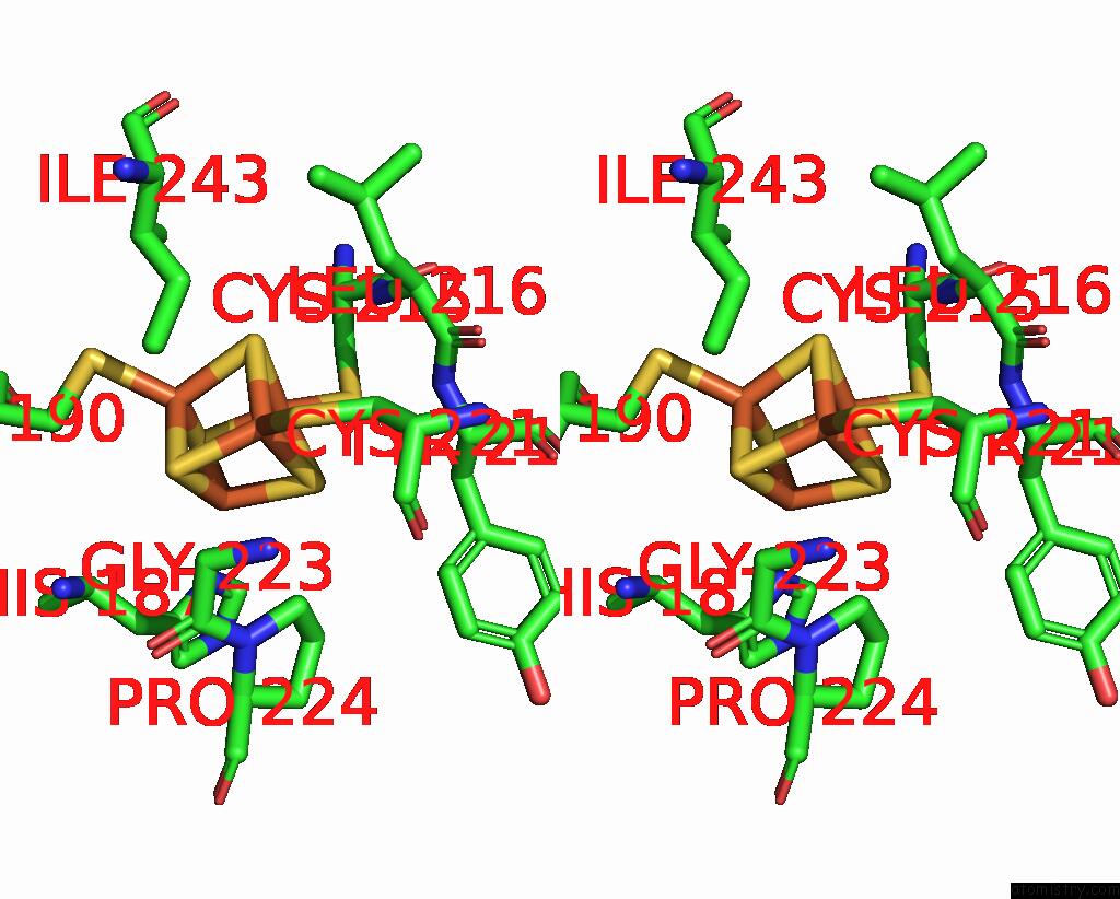
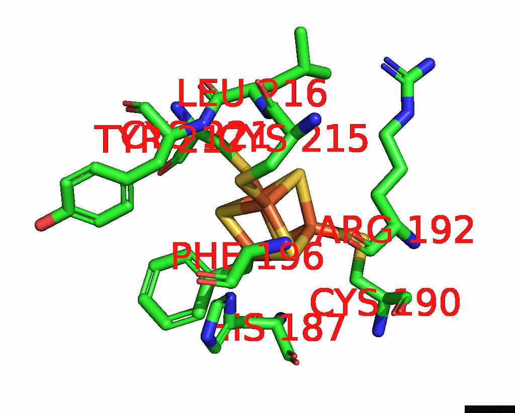
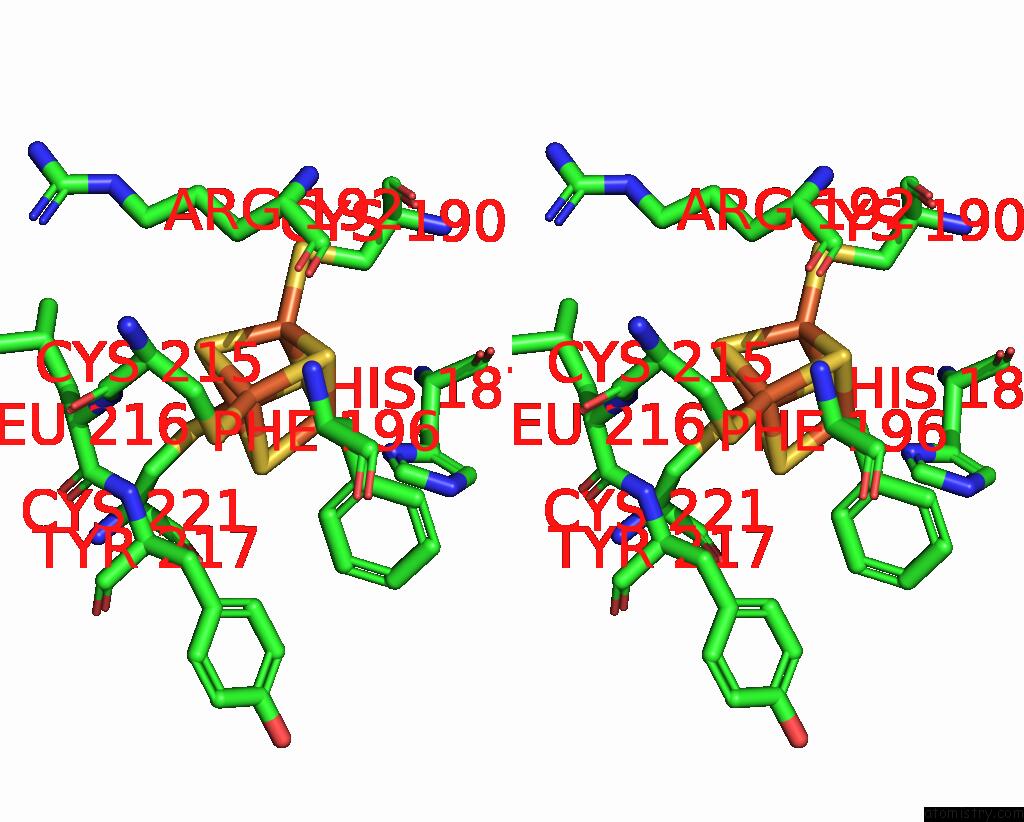
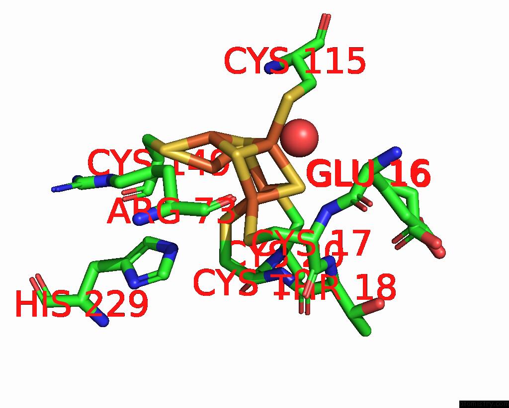
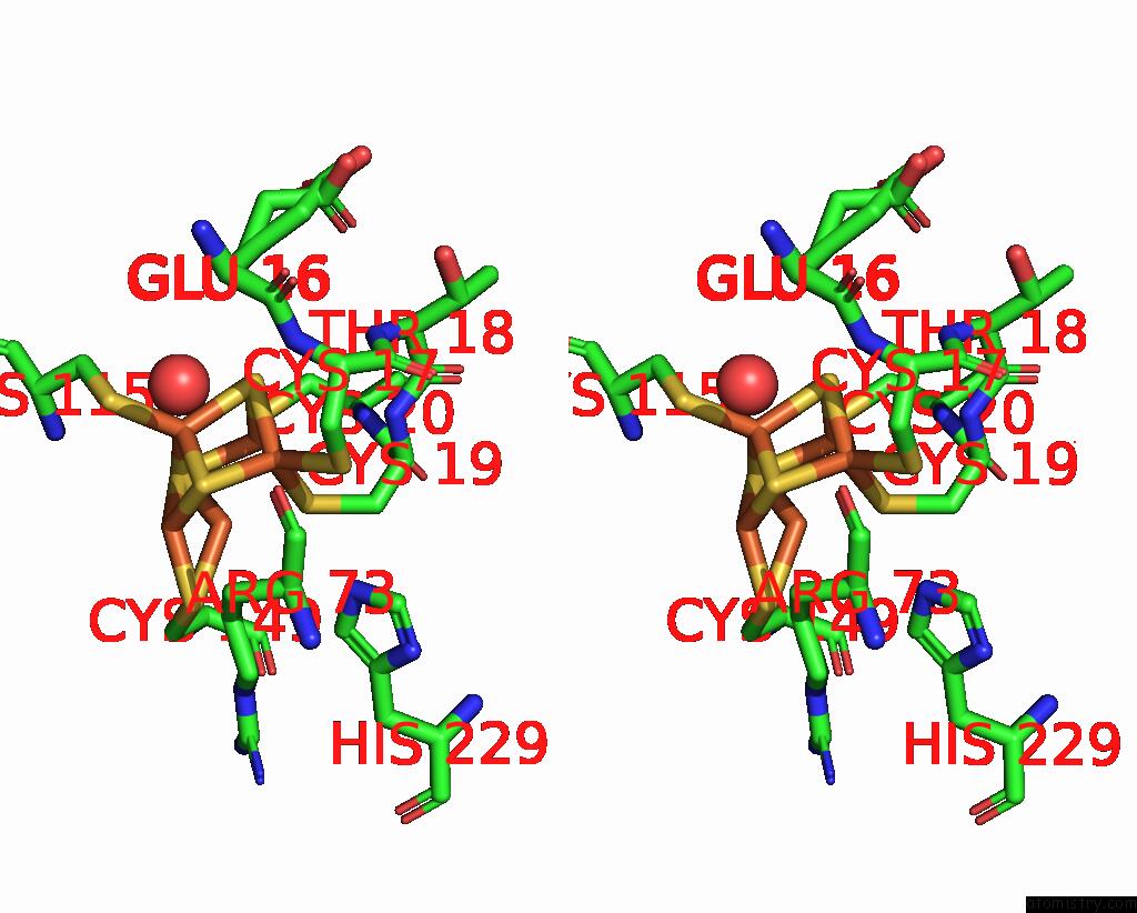
Iron binding site 1 out of 16 in 8poz
Go back to
Iron binding site 1 out
of 16 in the Crystal Structure of the C120G Variant of the Membrane-Bound [Nife]- Hydrogenase From Cupriavidus Necator in the H2-Reduced State at 1.65 A Resolution.
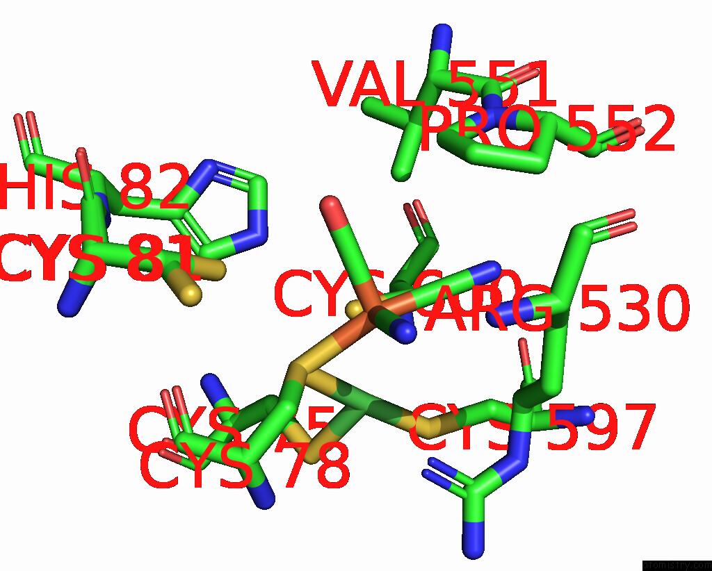
Mono view
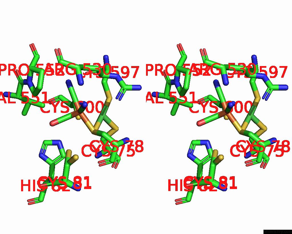
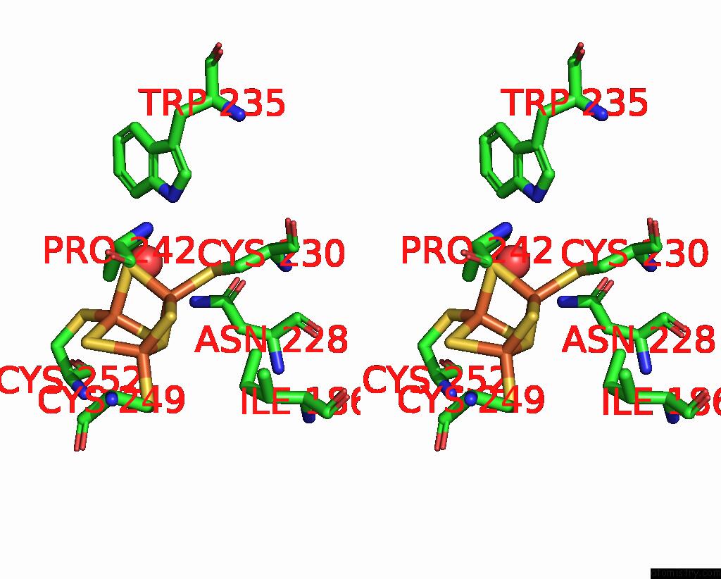
Stereo pair view
Mono view
Stereo pair view
A full contact list of Iron with other atoms in the Fe binding
site number 1 of Crystal Structure of the C120G Variant of the Membrane-Bound [Nife]- Hydrogenase From Cupriavidus Necator in the H2-Reduced State at 1.65 A Resolution. within 5.0Å range:
|
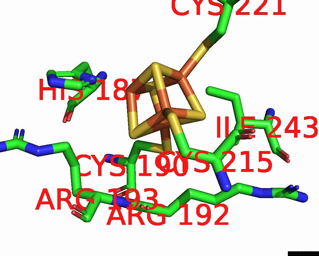
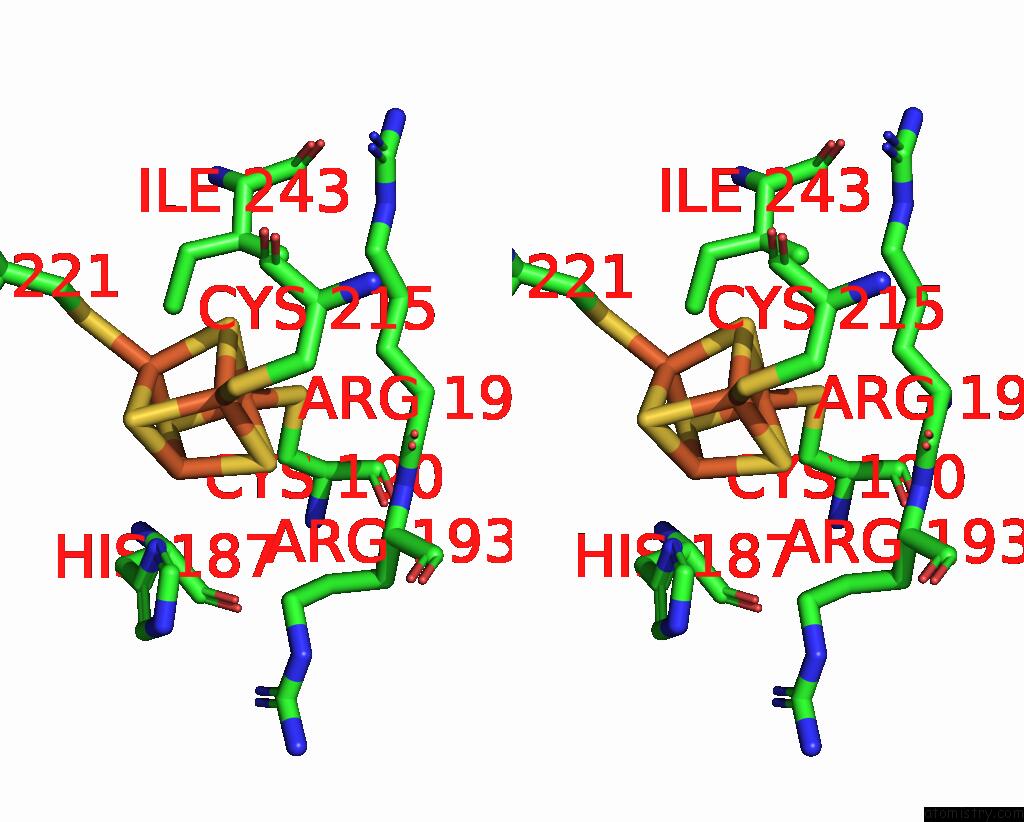
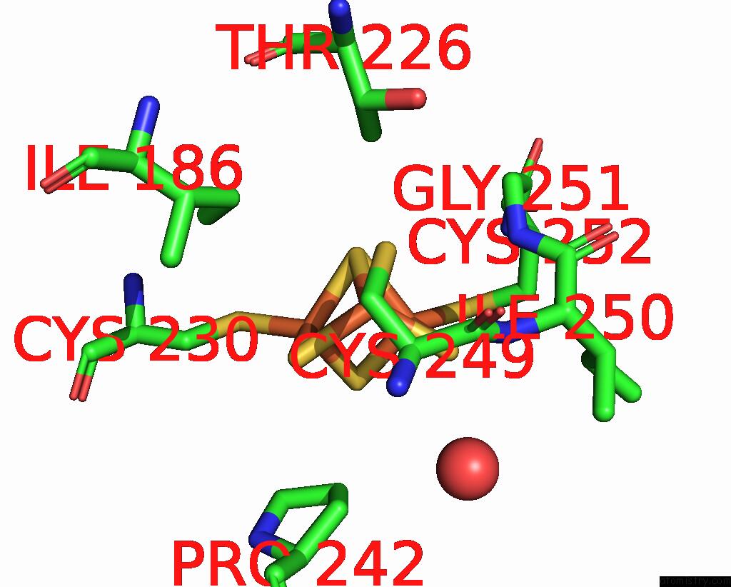
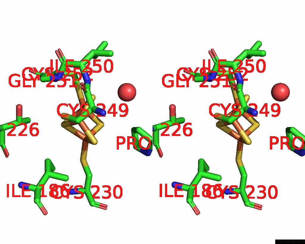
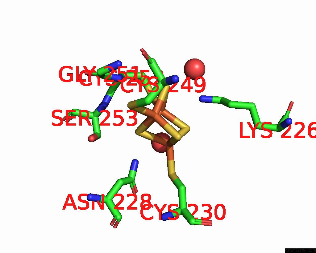
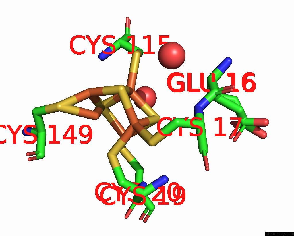
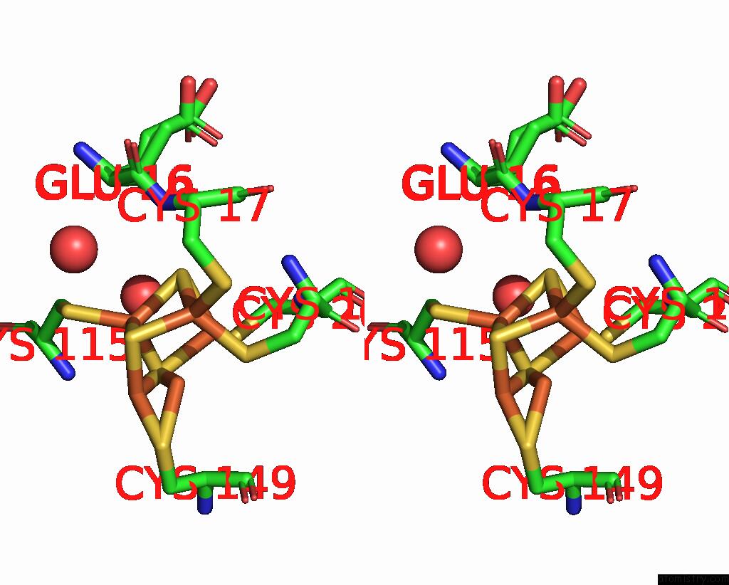
Iron binding site 2 out of 16 in 8poz
Go back to
Iron binding site 2 out
of 16 in the Crystal Structure of the C120G Variant of the Membrane-Bound [Nife]- Hydrogenase From Cupriavidus Necator in the H2-Reduced State at 1.65 A Resolution.
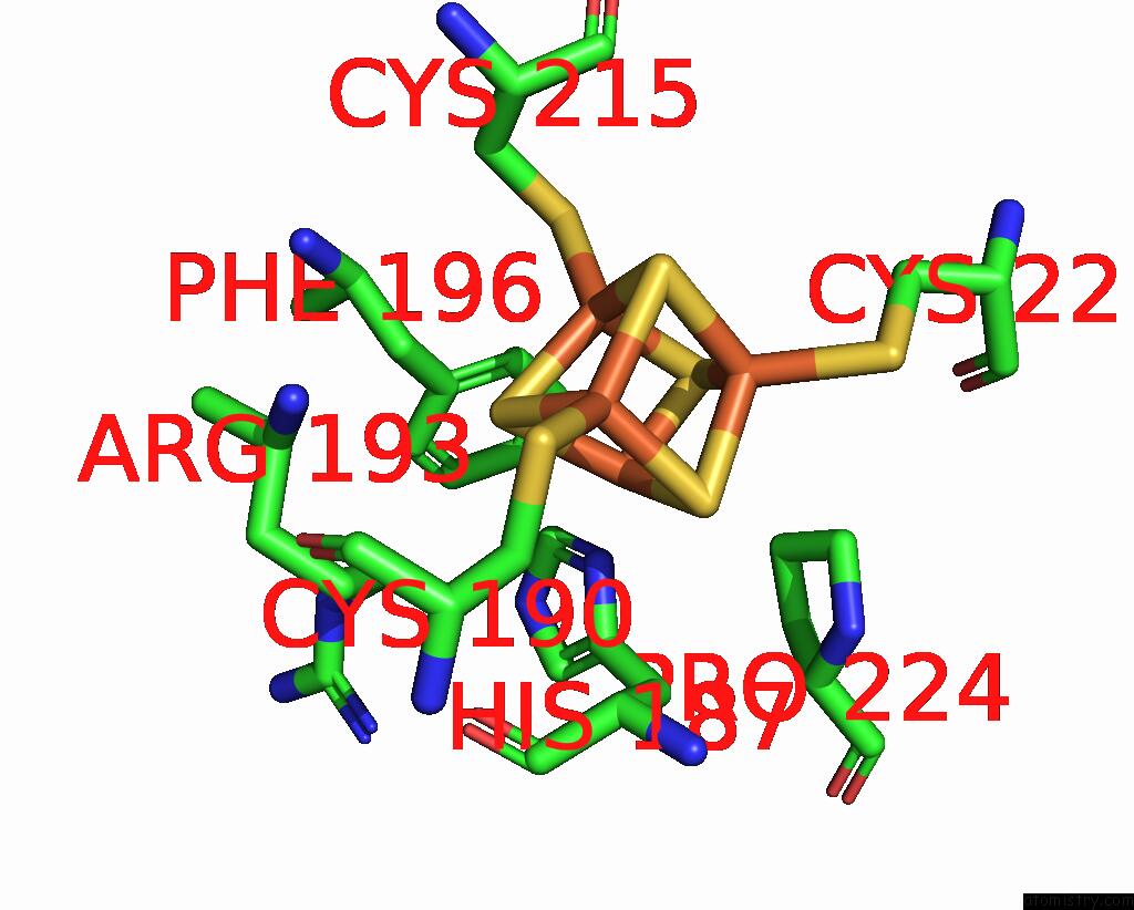
Mono view
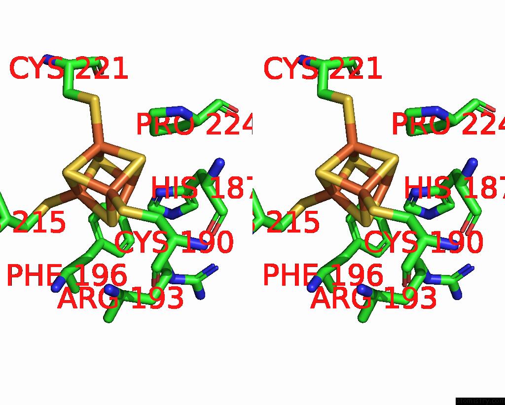
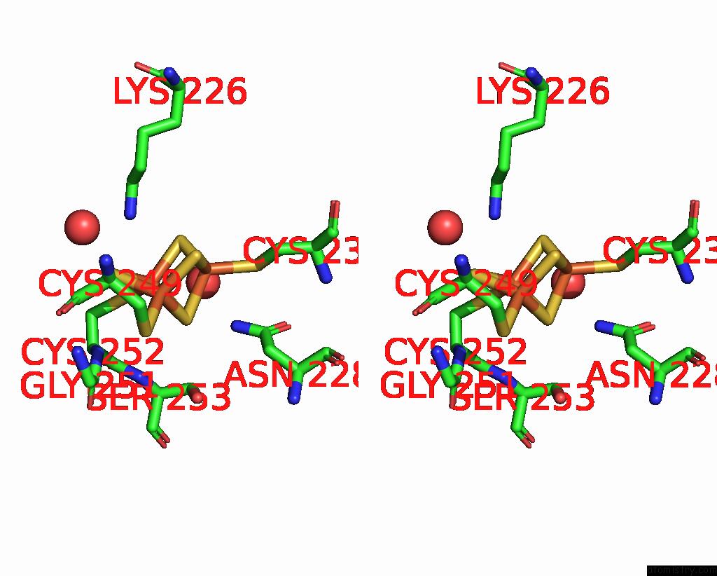
Stereo pair view
Mono view
Stereo pair view
A full contact list of Iron with other atoms in the Fe binding
site number 2 of Crystal Structure of the C120G Variant of the Membrane-Bound [Nife]- Hydrogenase From Cupriavidus Necator in the H2-Reduced State at 1.65 A Resolution. within 5.0Å range:
|
Iron binding site 3 out of 16 in 8poz
Go back to
Iron binding site 3 out
of 16 in the Crystal Structure of the C120G Variant of the Membrane-Bound [Nife]- Hydrogenase From Cupriavidus Necator in the H2-Reduced State at 1.65 A Resolution.
Mono view
Stereo pair view
Mono view
Stereo pair view
A full contact list of Iron with other atoms in the Fe binding
site number 3 of Crystal Structure of the C120G Variant of the Membrane-Bound [Nife]- Hydrogenase From Cupriavidus Necator in the H2-Reduced State at 1.65 A Resolution. within 5.0Å range:
|
Iron binding site 4 out of 16 in 8poz
Go back to
Iron binding site 4 out
of 16 in the Crystal Structure of the C120G Variant of the Membrane-Bound [Nife]- Hydrogenase From Cupriavidus Necator in the H2-Reduced State at 1.65 A Resolution.
Mono view
Stereo pair view
Mono view
Stereo pair view
A full contact list of Iron with other atoms in the Fe binding
site number 4 of Crystal Structure of the C120G Variant of the Membrane-Bound [Nife]- Hydrogenase From Cupriavidus Necator in the H2-Reduced State at 1.65 A Resolution. within 5.0Å range:
|
Iron binding site 5 out of 16 in 8poz
Go back to
Iron binding site 5 out
of 16 in the Crystal Structure of the C120G Variant of the Membrane-Bound [Nife]- Hydrogenase From Cupriavidus Necator in the H2-Reduced State at 1.65 A Resolution.
Mono view
Stereo pair view
Mono view
Stereo pair view
A full contact list of Iron with other atoms in the Fe binding
site number 5 of Crystal Structure of the C120G Variant of the Membrane-Bound [Nife]- Hydrogenase From Cupriavidus Necator in the H2-Reduced State at 1.65 A Resolution. within 5.0Å range:
|
Iron binding site 6 out of 16 in 8poz
Go back to
Iron binding site 6 out
of 16 in the Crystal Structure of the C120G Variant of the Membrane-Bound [Nife]- Hydrogenase From Cupriavidus Necator in the H2-Reduced State at 1.65 A Resolution.
Mono view
Stereo pair view
Mono view
Stereo pair view
A full contact list of Iron with other atoms in the Fe binding
site number 6 of Crystal Structure of the C120G Variant of the Membrane-Bound [Nife]- Hydrogenase From Cupriavidus Necator in the H2-Reduced State at 1.65 A Resolution. within 5.0Å range:
|
Iron binding site 7 out of 16 in 8poz
Go back to
Iron binding site 7 out
of 16 in the Crystal Structure of the C120G Variant of the Membrane-Bound [Nife]- Hydrogenase From Cupriavidus Necator in the H2-Reduced State at 1.65 A Resolution.
Mono view
Stereo pair view
Mono view
Stereo pair view
A full contact list of Iron with other atoms in the Fe binding
site number 7 of Crystal Structure of the C120G Variant of the Membrane-Bound [Nife]- Hydrogenase From Cupriavidus Necator in the H2-Reduced State at 1.65 A Resolution. within 5.0Å range:
|
Iron binding site 8 out of 16 in 8poz
Go back to
Iron binding site 8 out
of 16 in the Crystal Structure of the C120G Variant of the Membrane-Bound [Nife]- Hydrogenase From Cupriavidus Necator in the H2-Reduced State at 1.65 A Resolution.
Mono view
Stereo pair view
Mono view
Stereo pair view
A full contact list of Iron with other atoms in the Fe binding
site number 8 of Crystal Structure of the C120G Variant of the Membrane-Bound [Nife]- Hydrogenase From Cupriavidus Necator in the H2-Reduced State at 1.65 A Resolution. within 5.0Å range:
|
Iron binding site 9 out of 16 in 8poz
Go back to
Iron binding site 9 out
of 16 in the Crystal Structure of the C120G Variant of the Membrane-Bound [Nife]- Hydrogenase From Cupriavidus Necator in the H2-Reduced State at 1.65 A Resolution.
Mono view
Stereo pair view
Mono view
Stereo pair view
A full contact list of Iron with other atoms in the Fe binding
site number 9 of Crystal Structure of the C120G Variant of the Membrane-Bound [Nife]- Hydrogenase From Cupriavidus Necator in the H2-Reduced State at 1.65 A Resolution. within 5.0Å range:
|
Iron binding site 10 out of 16 in 8poz
Go back to
Iron binding site 10 out
of 16 in the Crystal Structure of the C120G Variant of the Membrane-Bound [Nife]- Hydrogenase From Cupriavidus Necator in the H2-Reduced State at 1.65 A Resolution.
Mono view
Stereo pair view
Mono view
Stereo pair view
A full contact list of Iron with other atoms in the Fe binding
site number 10 of Crystal Structure of the C120G Variant of the Membrane-Bound [Nife]- Hydrogenase From Cupriavidus Necator in the H2-Reduced State at 1.65 A Resolution. within 5.0Å range:
|
Reference:
A.Schmidt,
J.Kalms,
C.Lorent,
S.Katz,
S.Frielingsdorf,
R.M.Evans,
J.Fritsch,
E.Siebert,
C.Teutloff,
F.A.Armstrong,
I.Zebger,
O.Lenz,
P.Scheerer.
Stepwise Conversion of the Cys 6 [4FE-3S] to A Cys 4 [4FE-4S] Cluster and Its Impact on the Oxygen Tolerance of [Nife]-Hydrogenase. Chem Sci V. 14 11105 2023.
ISSN: ISSN 2041-6520
PubMed: 37860641
DOI: 10.1039/D3SC03739H
Page generated: Sat Sep 28 21:35:29 2024
ISSN: ISSN 2041-6520
PubMed: 37860641
DOI: 10.1039/D3SC03739H
Last articles
Fe in 2YXOFe in 2YRS
Fe in 2YXC
Fe in 2YNM
Fe in 2YVJ
Fe in 2YP1
Fe in 2YU2
Fe in 2YU1
Fe in 2YQB
Fe in 2YOO