Atomistry » Iron » PDB 8oz5-8pxq » 8pp5
Atomistry »
  Iron »
    PDB 8oz5-8pxq »
      8pp5 »

Iron in PDB 8pp5: Unitary Crystal Structure of Positively Supercharged Ferritin Variant Ftn(Pos)-M1 (Mg Formate Condition)

Protein crystallography data

The structure of Unitary Crystal Structure of Positively Supercharged Ferritin Variant Ftn(Pos)-M1 (Mg Formate Condition), PDB code: 8pp5 was solved by L.Lang, T.Beck, with X-Ray Crystallography technique. A brief refinement statistics is given in the table below:

Resolution Low / High (Å) 105.09 / 2.00
Space group I 4
Cell size a, b, c (Å), α, β, γ (°) 126.898, 126.898, 187.468, 90, 90, 90
R / Rfree (%) 16 / 19.3

Other elements in 8pp5:

The structure of Unitary Crystal Structure of Positively Supercharged Ferritin Variant Ftn(Pos)-M1 (Mg Formate Condition) also contains other interesting chemical elements:

Magnesium (Mg) 4 atoms

Iron Binding Sites:

The binding sites of Iron atom in the Unitary Crystal Structure of Positively Supercharged Ferritin Variant Ftn(Pos)-M1 (Mg Formate Condition) (pdb code 8pp5). This binding sites where shown within 5.0 Angstroms radius around Iron atom.
In total 6 binding sites of Iron where determined in the Unitary Crystal Structure of Positively Supercharged Ferritin Variant Ftn(Pos)-M1 (Mg Formate Condition), PDB code: 8pp5:
Jump to Iron binding site number: 1; 2; 3; 4; 5; 6;

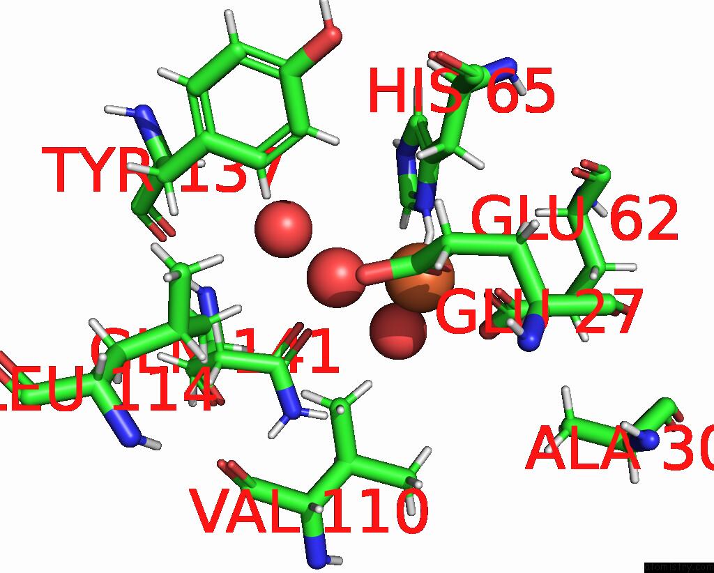
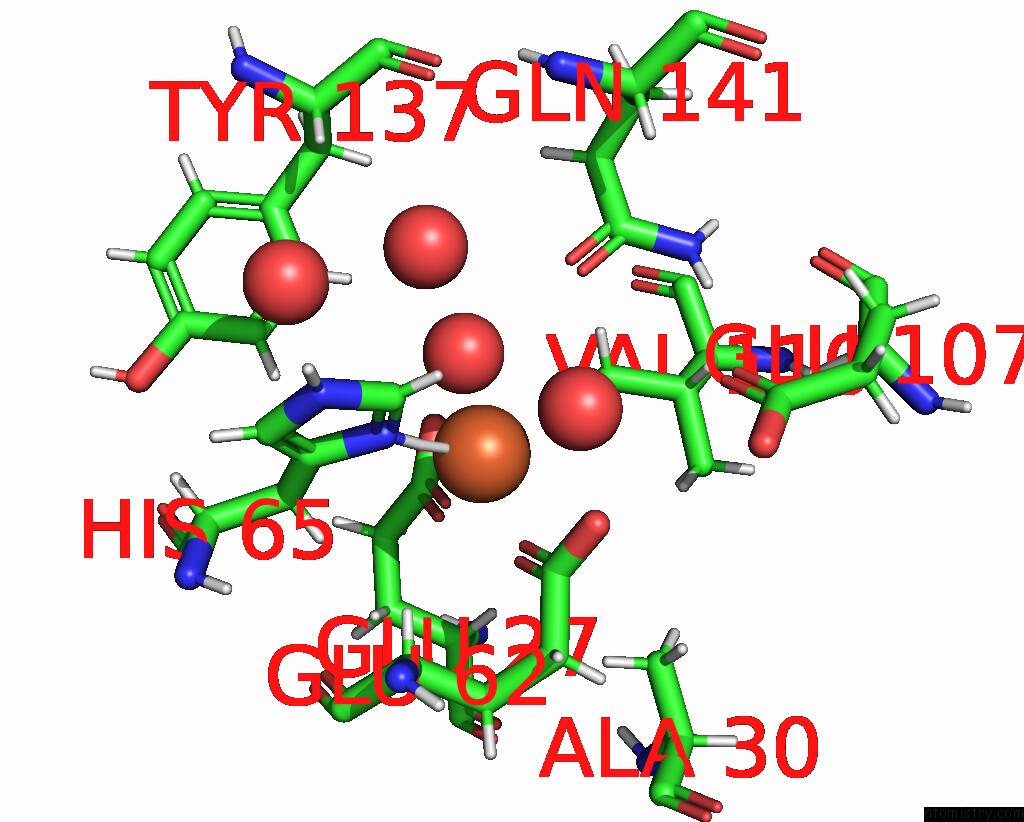
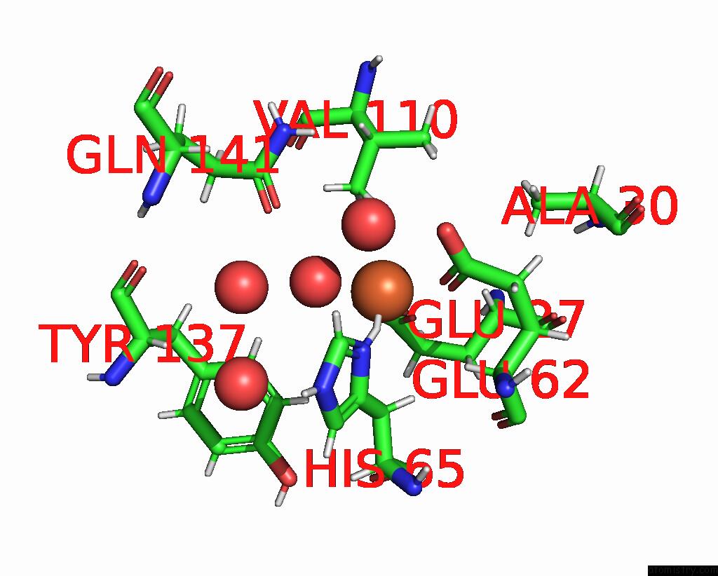
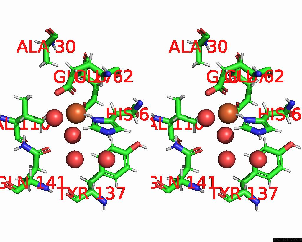
Iron binding site 1 out of 6 in 8pp5

Go back to Iron Binding Sites List in 8pp5
Iron binding site 1 out of 6 in the Unitary Crystal Structure of Positively Supercharged Ferritin Variant Ftn(Pos)-M1 (Mg Formate Condition)


Mono view


Stereo pair view

A full contact list of Iron with other atoms in the Fe binding site number 1 of Unitary Crystal Structure of Positively Supercharged Ferritin Variant Ftn(Pos)-M1 (Mg Formate Condition) within 5.0Å range:
probe atom residue distance (Å) B Occ
A:Fe201

b:41.4
occ:1.00
HD1 A:HIS65 1.4 31.4 0.0
OE1 A:GLU62 2.0 30.7 1.0
OE1 A:GLU27 2.1 27.4 1.0
O A:HOH317 2.2 34.1 1.0
ND1 A:HIS65 2.2 31.7 1.0
O A:HOH305 2.2 27.6 1.0
CD A:GLU62 3.0 30.8 1.0
CD A:GLU27 3.1 25.8 1.0
CE1 A:HIS65 3.2 34.1 1.0
CG A:HIS65 3.3 28.8 1.0
HE1 A:HIS65 3.3 33.1 1.0
OE2 A:GLU62 3.3 34.0 1.0
HG13 A:VAL110 3.4 24.7 1.0
OE2 A:GLU27 3.4 27.6 1.0
HB2 A:HIS65 3.4 24.8 1.0
HB3 A:HIS65 3.4 24.8 1.0
CB A:HIS65 3.6 24.1 1.0
HA A:GLU62 3.7 22.7 1.0
OE1 A:GLN141 4.0 34.6 1.0
O A:HOH390 4.0 34.2 1.0
HA A:GLU27 4.3 21.7 1.0
CG1 A:VAL110 4.3 25.1 1.0
NE2 A:HIS65 4.3 32.3 1.0
CG A:GLU62 4.3 24.8 1.0
CD2 A:HIS65 4.4 30.4 1.0
HB3 A:GLU27 4.4 21.3 1.0
CG A:GLU27 4.4 21.7 1.0
HG12 A:VAL110 4.4 24.6 1.0
HB3 A:GLU62 4.5 23.3 1.0
HG21 A:VAL110 4.5 25.6 1.0
CA A:GLU62 4.5 23.5 1.0
HE2 A:TYR137 4.6 24.8 1.0
HD2 A:TYR137 4.6 25.4 1.0
CB A:GLU62 4.7 22.6 1.0
HG11 A:VAL110 4.8 24.6 1.0
HB3 A:ALA30 4.8 24.6 1.0
CB A:GLU27 4.8 20.7 1.0
HG3 A:GLU27 4.8 22.3 1.0
HG3 A:GLU62 4.9 25.5 1.0
HG2 A:GLU62 4.9 25.5 1.0
CD A:GLN141 4.9 33.1 1.0
HD11 A:LEU114 5.0 24.3 1.0

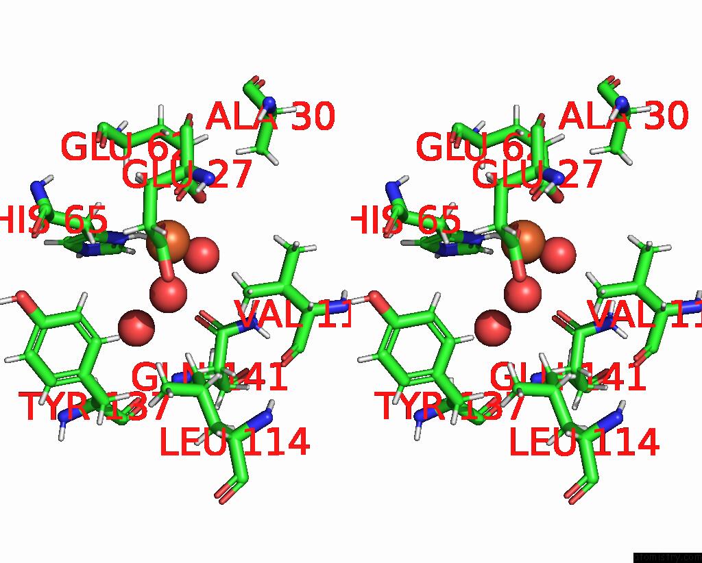
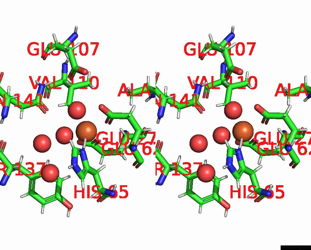
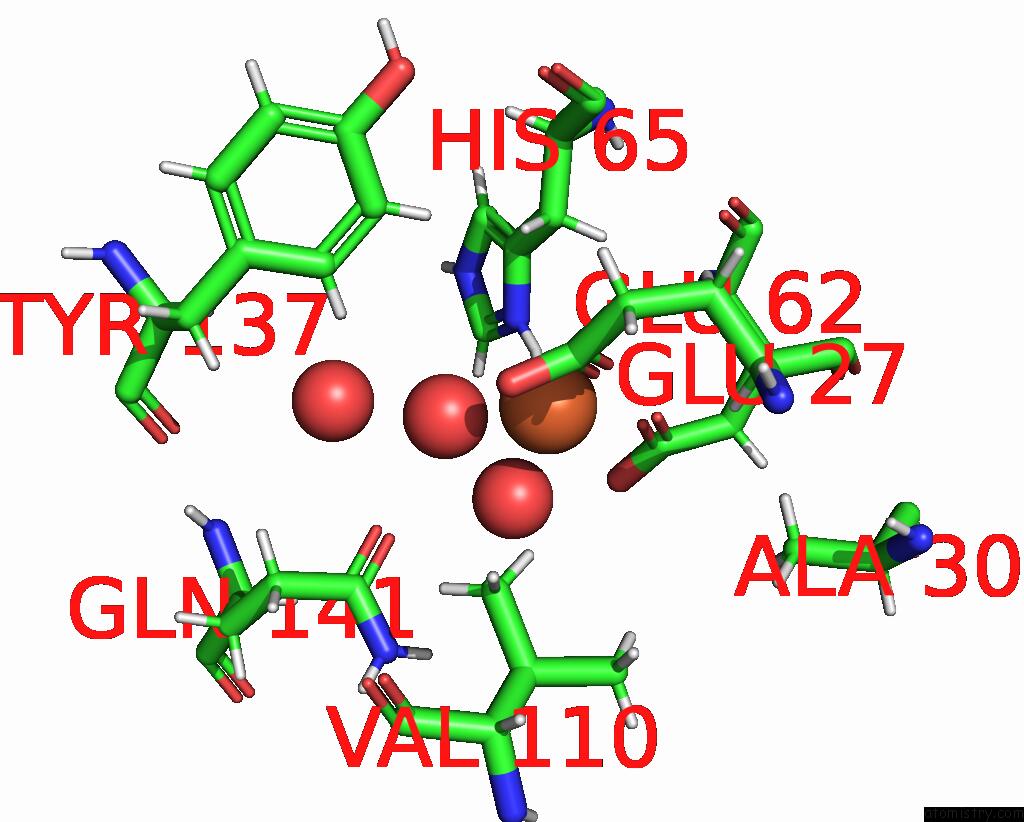
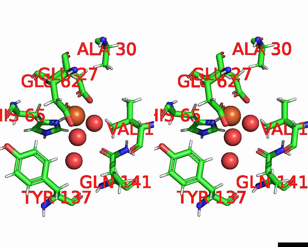
Iron binding site 2 out of 6 in 8pp5

Go back to Iron Binding Sites List in 8pp5
Iron binding site 2 out of 6 in the Unitary Crystal Structure of Positively Supercharged Ferritin Variant Ftn(Pos)-M1 (Mg Formate Condition)


Mono view


Stereo pair view

A full contact list of Iron with other atoms in the Fe binding site number 2 of Unitary Crystal Structure of Positively Supercharged Ferritin Variant Ftn(Pos)-M1 (Mg Formate Condition) within 5.0Å range:
probe atom residue distance (Å) B Occ
B:Fe201

b:42.8
occ:1.00
HD1 B:HIS65 1.4 26.9 0.0
OE1 B:GLU62 2.1 33.9 1.0
OE1 B:GLU27 2.1 29.0 1.0
O B:HOH317 2.2 34.5 1.0
ND1 B:HIS65 2.2 26.9 1.0
O B:HOH307 2.3 24.3 1.0
CD B:GLU62 3.0 28.1 1.0
CE1 B:HIS65 3.1 31.2 1.0
CD B:GLU27 3.1 26.4 1.0
HE1 B:HIS65 3.2 29.7 1.0
OE2 B:GLU62 3.3 35.8 1.0
HG13 B:VAL110 3.3 25.6 1.0
CG B:HIS65 3.3 28.5 1.0
OE2 B:GLU27 3.4 28.5 1.0
HB2 B:HIS65 3.5 24.9 1.0
HB3 B:HIS65 3.6 24.9 1.0
HA B:GLU62 3.7 23.7 1.0
CB B:HIS65 3.7 24.2 1.0
OE1 B:GLN141 4.0 34.8 1.0
O B:HOH368 4.0 38.8 1.0
CG1 B:VAL110 4.2 26.4 1.0
NE2 B:HIS65 4.2 29.1 1.0
HB3 B:GLU27 4.2 22.7 1.0
HA B:GLU27 4.3 22.2 1.0
HG12 B:VAL110 4.3 25.6 1.0
O B:HOH302 4.3 52.1 1.0
CG B:GLU62 4.4 22.8 1.0
CD2 B:HIS65 4.4 26.6 1.0
CG B:GLU27 4.4 25.9 1.0
HB3 B:GLU62 4.5 23.1 1.0
HG21 B:VAL110 4.5 25.2 1.0
CA B:GLU62 4.6 23.8 1.0
HD2 B:TYR137 4.6 21.4 1.0
HE2 B:TYR137 4.6 21.3 1.0
HG11 B:VAL110 4.7 25.6 1.0
CB B:GLU27 4.7 21.8 1.0
CB B:GLU62 4.7 22.9 1.0
HB3 B:ALA30 4.7 26.1 1.0
HG3 B:GLU27 4.8 25.0 1.0
OE1 B:GLU107 4.9 44.6 1.0
HG3 B:GLU62 4.9 24.0 1.0
CD B:GLN141 4.9 28.0 1.0
HG2 B:GLU62 4.9 23.9 1.0

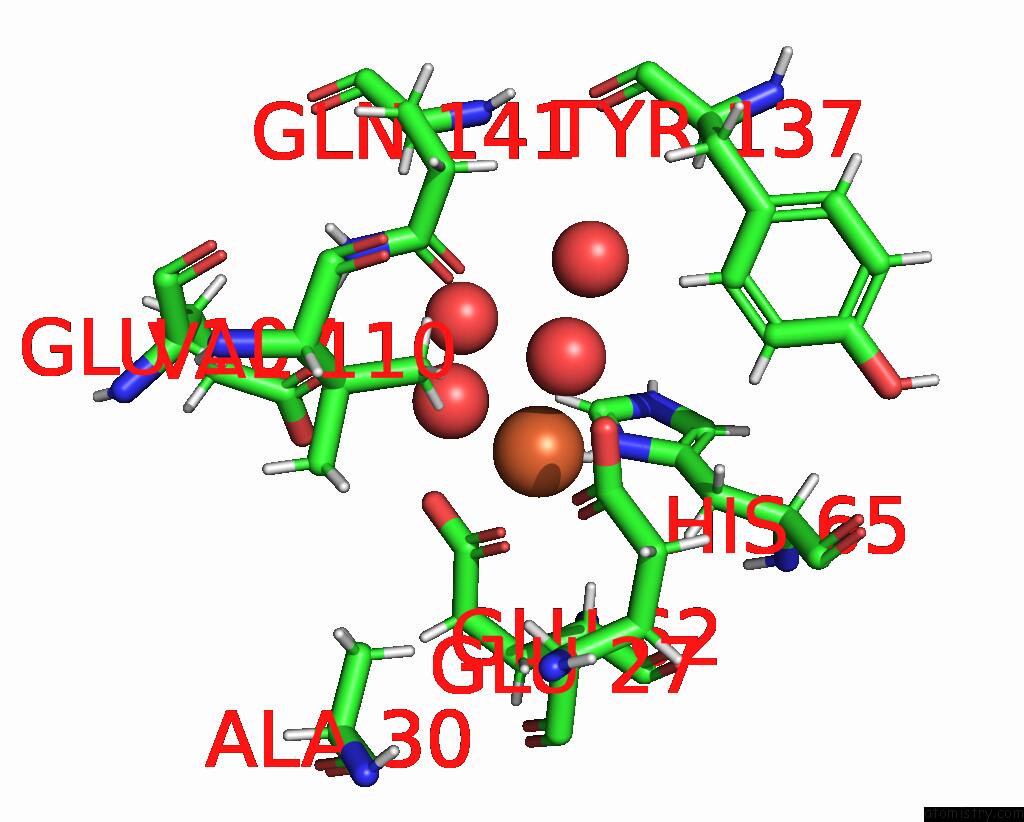
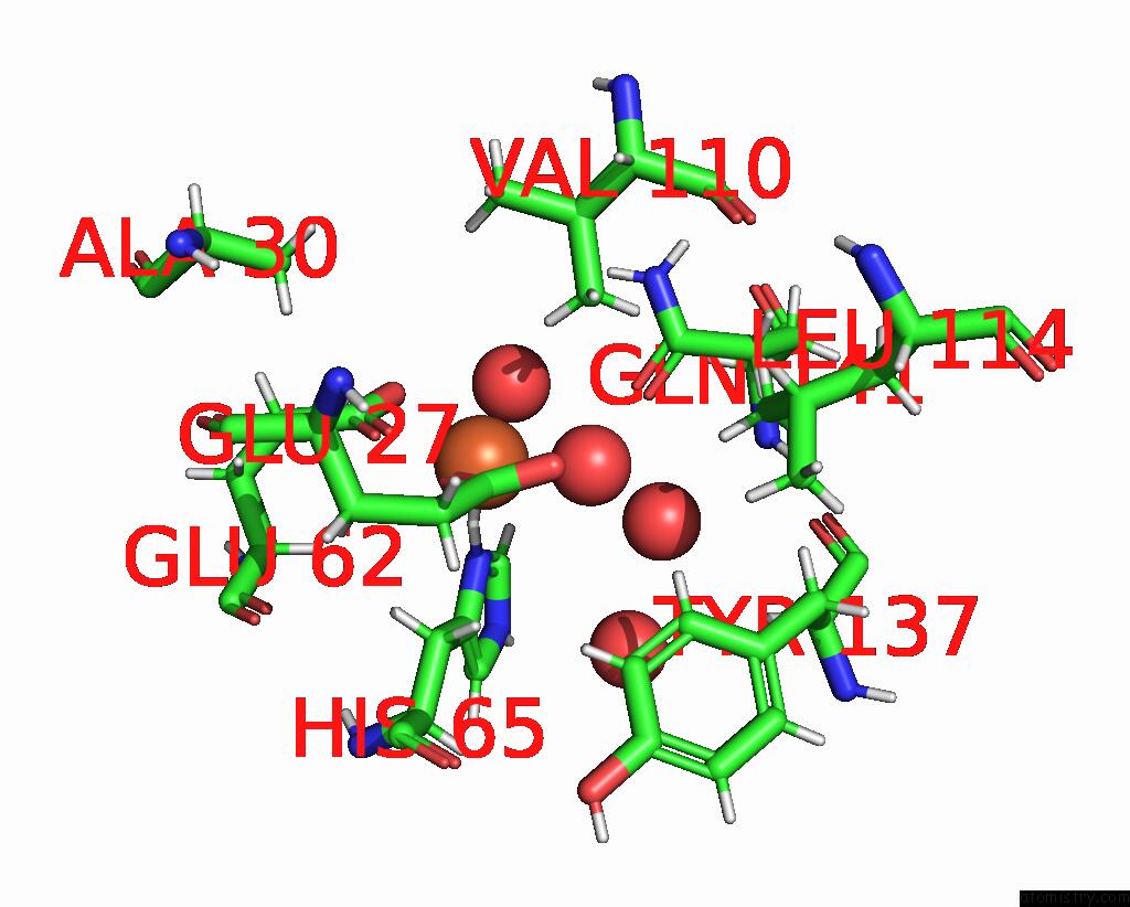
Iron binding site 3 out of 6 in 8pp5

Go back to Iron Binding Sites List in 8pp5
Iron binding site 3 out of 6 in the Unitary Crystal Structure of Positively Supercharged Ferritin Variant Ftn(Pos)-M1 (Mg Formate Condition)


Mono view


Stereo pair view

A full contact list of Iron with other atoms in the Fe binding site number 3 of Unitary Crystal Structure of Positively Supercharged Ferritin Variant Ftn(Pos)-M1 (Mg Formate Condition) within 5.0Å range:
probe atom residue distance (Å) B Occ
C:Fe201

b:43.5
occ:1.00
HD1 C:HIS65 1.5 32.4 0.0
OE1 C:GLU62 2.1 32.6 1.0
OE1 C:GLU27 2.1 28.1 1.0
O C:HOH325 2.1 38.3 1.0
O C:HOH318 2.2 27.1 1.0
ND1 C:HIS65 2.4 32.6 1.0
CD C:GLU62 3.0 29.0 1.0
CD C:GLU27 3.0 27.2 1.0
OE2 C:GLU27 3.3 26.7 1.0
OE2 C:GLU62 3.3 32.4 1.0
CE1 C:HIS65 3.3 39.6 1.0
HB2 C:HIS65 3.4 27.2 1.0
CG C:HIS65 3.4 31.4 1.0
HE1 C:HIS65 3.4 36.9 1.0
HG13 C:VAL110 3.5 29.4 1.0
HB3 C:HIS65 3.5 27.1 1.0
CB C:HIS65 3.6 26.7 1.0
HA C:GLU62 3.7 23.5 1.0
OE1 C:GLN141 3.9 35.3 1.0
O C:HOH382 4.0 42.5 1.0
HA C:GLU27 4.3 22.3 1.0
HB3 C:GLU27 4.3 23.5 1.0
CG1 C:VAL110 4.4 29.6 1.0
CG C:GLU27 4.4 23.4 1.0
CG C:GLU62 4.4 25.4 1.0
HG12 C:VAL110 4.4 29.4 1.0
NE2 C:HIS65 4.5 35.8 1.0
CD2 C:HIS65 4.5 31.4 1.0
HE2 C:TYR137 4.5 24.4 1.0
HB3 C:GLU62 4.5 24.7 1.0
HD2 C:TYR137 4.6 24.7 1.0
CA C:GLU62 4.6 23.7 1.0
HB3 C:ALA30 4.7 24.4 1.0
HG21 C:VAL110 4.7 27.5 1.0
CB C:GLU62 4.7 24.9 1.0
HG3 C:GLU27 4.7 24.4 1.0
CB C:GLU27 4.8 23.9 1.0
HG11 C:VAL110 4.9 29.4 1.0
O C:HOH401 4.9 42.9 1.0
HG3 C:GLU62 4.9 26.1 1.0
OE1 C:GLU107 4.9 39.5 1.0
CD C:GLN141 4.9 28.8 1.0
HG2 C:GLU62 4.9 26.1 1.0

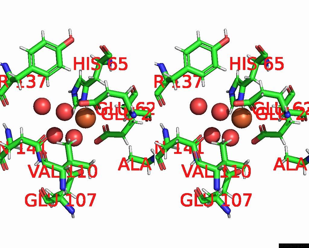
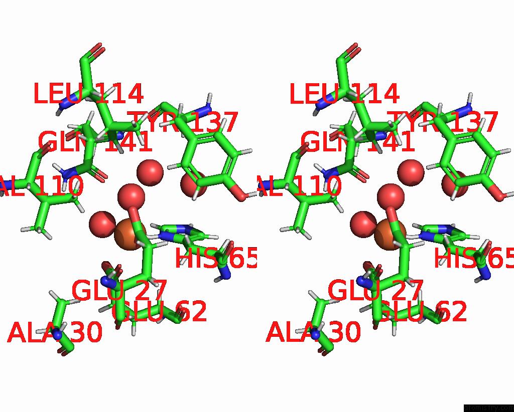
Iron binding site 4 out of 6 in 8pp5

Go back to Iron Binding Sites List in 8pp5
Iron binding site 4 out of 6 in the Unitary Crystal Structure of Positively Supercharged Ferritin Variant Ftn(Pos)-M1 (Mg Formate Condition)


Mono view


Stereo pair view

A full contact list of Iron with other atoms in the Fe binding site number 4 of Unitary Crystal Structure of Positively Supercharged Ferritin Variant Ftn(Pos)-M1 (Mg Formate Condition) within 5.0Å range:
probe atom residue distance (Å) B Occ
D:Fe201

b:47.9
occ:1.00
HD1 D:HIS65 1.3 33.8 0.0
OE1 D:GLU62 2.0 35.8 1.0
OE1 D:GLU27 2.1 29.8 1.0
ND1 D:HIS65 2.2 34.3 1.0
O D:HOH307 2.2 27.3 1.0
O D:HOH313 2.2 32.1 1.0
CD D:GLU62 3.0 32.4 1.0
CE1 D:HIS65 3.1 34.4 1.0
CD D:GLU27 3.1 25.6 1.0
HE1 D:HIS65 3.2 34.2 1.0
OE2 D:GLU62 3.2 37.8 1.0
CG D:HIS65 3.2 32.6 1.0
HG13 D:VAL110 3.3 29.1 1.0
OE2 D:GLU27 3.4 28.0 1.0
HB2 D:HIS65 3.4 27.0 1.0
HB3 D:HIS65 3.4 26.9 1.0
CB D:HIS65 3.6 25.9 1.0
HA D:GLU62 3.7 24.4 1.0
O D:HOH381 3.9 38.9 1.0
OE1 D:GLN141 4.1 33.5 1.0
CG1 D:VAL110 4.2 30.6 1.0
NE2 D:HIS65 4.2 33.7 1.0
HG12 D:VAL110 4.3 29.1 1.0
HB3 D:GLU27 4.3 24.5 1.0
HA D:GLU27 4.3 23.8 1.0
CG D:GLU62 4.3 26.1 1.0
CD2 D:HIS65 4.3 32.8 1.0
CG D:GLU27 4.4 25.3 1.0
HD2 D:TYR137 4.5 24.1 1.0
HE2 D:TYR137 4.6 24.0 1.0
HG11 D:VAL110 4.6 29.1 1.0
HG21 D:VAL110 4.6 26.2 1.0
CA D:GLU62 4.6 24.6 1.0
HB3 D:GLU62 4.6 23.9 1.0
CB D:GLU27 4.7 24.4 1.0
CB D:GLU62 4.8 22.7 1.0
HB3 D:ALA30 4.8 22.6 1.0
HG3 D:GLU27 4.8 25.2 1.0
HG3 D:GLU62 4.8 26.6 1.0
HG2 D:GLU62 4.9 26.6 1.0
O D:HOH389 4.9 45.6 1.0
HD11 D:LEU114 4.9 24.3 1.0
CD D:GLN141 5.0 30.7 1.0
HE2 D:HIS65 5.0 33.0 0.0

Iron binding site 5 out of 6 in 8pp5

Go back to Iron Binding Sites List in 8pp5
Iron binding site 5 out of 6 in the Unitary Crystal Structure of Positively Supercharged Ferritin Variant Ftn(Pos)-M1 (Mg Formate Condition)


Mono view


Stereo pair view

A full contact list of Iron with other atoms in the Fe binding site number 5 of Unitary Crystal Structure of Positively Supercharged Ferritin Variant Ftn(Pos)-M1 (Mg Formate Condition) within 5.0Å range:
probe atom residue distance (Å) B Occ
E:Fe201

b:44.9
occ:1.00
HD1 E:HIS65 1.3 28.0 0.0
OE1 E:GLU62 2.0 29.6 1.0
OE1 E:GLU27 2.1 26.1 1.0
O E:HOH365 2.1 39.7 1.0
ND1 E:HIS65 2.2 28.3 1.0
O E:HOH333 2.3 29.9 1.0
CD E:GLU62 3.0 28.9 1.0
CD E:GLU27 3.1 24.6 1.0
CE1 E:HIS65 3.1 34.7 1.0
HE1 E:HIS65 3.2 33.4 1.0
CG E:HIS65 3.2 31.5 1.0
HB2 E:HIS65 3.3 26.4 1.0
OE2 E:GLU27 3.4 28.3 1.0
HG13 E:VAL110 3.4 28.7 1.0
OE2 E:GLU62 3.4 37.9 1.0
HB3 E:HIS65 3.4 26.4 1.0
CB E:HIS65 3.6 26.2 1.0
HA E:GLU62 3.6 23.2 1.0
OE1 E:GLN141 4.1 40.4 1.0
O E:HOH309 4.1 35.3 1.0
NE2 E:HIS65 4.2 36.3 1.0
HA E:GLU27 4.3 23.1 1.0
CG1 E:VAL110 4.3 29.1 1.0
HB3 E:GLU27 4.3 22.6 1.0
HG12 E:VAL110 4.3 28.7 1.0
CG E:GLU62 4.3 25.1 1.0
CD2 E:HIS65 4.3 31.2 1.0
CG E:GLU27 4.4 21.1 1.0
HB3 E:GLU62 4.4 23.4 1.0
HD2 E:TYR137 4.5 25.2 1.0
CA E:GLU62 4.5 23.5 1.0
HE2 E:TYR137 4.6 24.1 1.0
CB E:GLU62 4.7 22.7 1.0
HB3 E:ALA30 4.7 23.5 1.0
HG3 E:GLU27 4.7 22.3 1.0
CB E:GLU27 4.7 22.6 1.0
HG21 E:VAL110 4.7 29.3 1.0
HG11 E:VAL110 4.8 28.7 1.0
HG2 E:GLU62 4.9 25.3 1.0
O E:HOH391 4.9 48.3 1.0
HG3 E:GLU62 4.9 25.4 1.0
CD E:GLN141 5.0 28.9 1.0

Iron binding site 6 out of 6 in 8pp5

Go back to Iron Binding Sites List in 8pp5
Iron binding site 6 out of 6 in the Unitary Crystal Structure of Positively Supercharged Ferritin Variant Ftn(Pos)-M1 (Mg Formate Condition)


Mono view


Stereo pair view

A full contact list of Iron with other atoms in the Fe binding site number 6 of Unitary Crystal Structure of Positively Supercharged Ferritin Variant Ftn(Pos)-M1 (Mg Formate Condition) within 5.0Å range:
probe atom residue distance (Å) B Occ
F:Fe201

b:47.4
occ:1.00
HD1 F:HIS65 1.4 29.8 0.0
OE1 F:GLU27 2.1 27.6 1.0
OE1 F:GLU62 2.1 30.1 1.0
O F:HOH315 2.2 31.1 1.0
O F:HOH305 2.2 28.9 1.0
ND1 F:HIS65 2.2 29.7 1.0
CD F:GLU27 3.0 27.6 1.0
CD F:GLU62 3.1 28.5 1.0
CE1 F:HIS65 3.1 32.0 1.0
CG F:HIS65 3.2 27.9 1.0
HE1 F:HIS65 3.3 31.0 1.0
HB2 F:HIS65 3.3 27.0 1.0
OE2 F:GLU27 3.3 27.2 1.0
OE2 F:GLU62 3.4 36.7 1.0
HB3 F:HIS65 3.4 27.0 1.0
HG13 F:VAL110 3.4 29.6 1.0
CB F:HIS65 3.5 27.6 1.0
HA F:GLU62 3.7 23.8 1.0
O F:HOH371 4.1 37.5 1.0
OE1 F:GLN141 4.1 35.4 1.0
HA F:GLU27 4.2 23.2 1.0
CG1 F:VAL110 4.3 30.5 1.0
NE2 F:HIS65 4.3 30.4 1.0
HB3 F:GLU27 4.3 24.0 1.0
CG F:GLU27 4.3 25.9 1.0
HG12 F:VAL110 4.3 29.6 1.0
CD2 F:HIS65 4.4 30.9 1.0
CG F:GLU62 4.4 25.8 1.0
HD2 F:TYR137 4.5 24.8 1.0
HB3 F:GLU62 4.5 23.6 1.0
HE2 F:TYR137 4.5 23.5 1.0
CA F:GLU62 4.6 23.9 1.0
HG11 F:VAL110 4.7 29.6 1.0
CB F:GLU27 4.7 23.3 1.0
HG3 F:GLU27 4.7 25.6 1.0
HB3 F:ALA30 4.7 24.1 1.0
CB F:GLU62 4.7 22.5 1.0
HG21 F:VAL110 4.7 27.4 1.0
HG3 F:GLU62 4.9 25.6 1.0
HG2 F:GLU62 4.9 25.5 1.0
HG2 F:GLU27 5.0 25.6 1.0
CA F:GLU27 5.0 24.1 1.0

Reference:

L.Lang, H.Bohler, H.Wagler, T.Beck. Assembly Requirements For the Construction of Large-Scale Binary Protein Structures. Biomacromolecules 2023.
ISSN: ESSN 1526-4602
PubMed: 38059469
DOI: 10.1021/ACS.BIOMAC.3C00891
Page generated: Sat Aug 10 13:08:50 2024

Last articles

Ca in 5NMR
Ca in 5NN9
Ca in 5NM8
Ca in 5NH8
Ca in 5NL7
Ca in 5NIN
Ca in 5NGQ
Ca in 5NH5
Ca in 5NGY
Ca in 5NG1
© Copyright 2008-2020 by atomistry.com
Home   |    Site Map   |    Copyright   |    Contact us   |    Privacy