Iron »
PDB 8qvb-8r9i »
8r2v »
Iron in PDB 8r2v: Crystal Structure of Hydroxyquinol-1,2-Dioxygenase From Gelatoporia Subvermispora (GSHDX1)
Protein crystallography data
The structure of Crystal Structure of Hydroxyquinol-1,2-Dioxygenase From Gelatoporia Subvermispora (GSHDX1), PDB code: 8r2v
was solved by
M.Zahn,
E.Kuatsjah,
D.Salvachua,
with X-Ray Crystallography technique. A brief refinement statistics is given in the table below:
| Resolution Low / High (Å) | 108.19 / 1.78 |
| Space group | P 21 21 21 |
| Cell size a, b, c (Å), α, β, γ (°) | 48.433, 148.662, 157.746, 90, 90, 90 |
| R / Rfree (%) | 17.3 / 19.7 |
Iron Binding Sites:
The binding sites of Iron atom in the Crystal Structure of Hydroxyquinol-1,2-Dioxygenase From Gelatoporia Subvermispora (GSHDX1)
(pdb code 8r2v). This binding sites where shown within
5.0 Angstroms radius around Iron atom.
In total 2 binding sites of Iron where determined in the Crystal Structure of Hydroxyquinol-1,2-Dioxygenase From Gelatoporia Subvermispora (GSHDX1), PDB code: 8r2v:
Jump to Iron binding site number: 1; 2;
In total 2 binding sites of Iron where determined in the Crystal Structure of Hydroxyquinol-1,2-Dioxygenase From Gelatoporia Subvermispora (GSHDX1), PDB code: 8r2v:
Jump to Iron binding site number: 1; 2;
Iron binding site 1 out of 2 in 8r2v
Go back to
Iron binding site 1 out
of 2 in the Crystal Structure of Hydroxyquinol-1,2-Dioxygenase From Gelatoporia Subvermispora (GSHDX1)
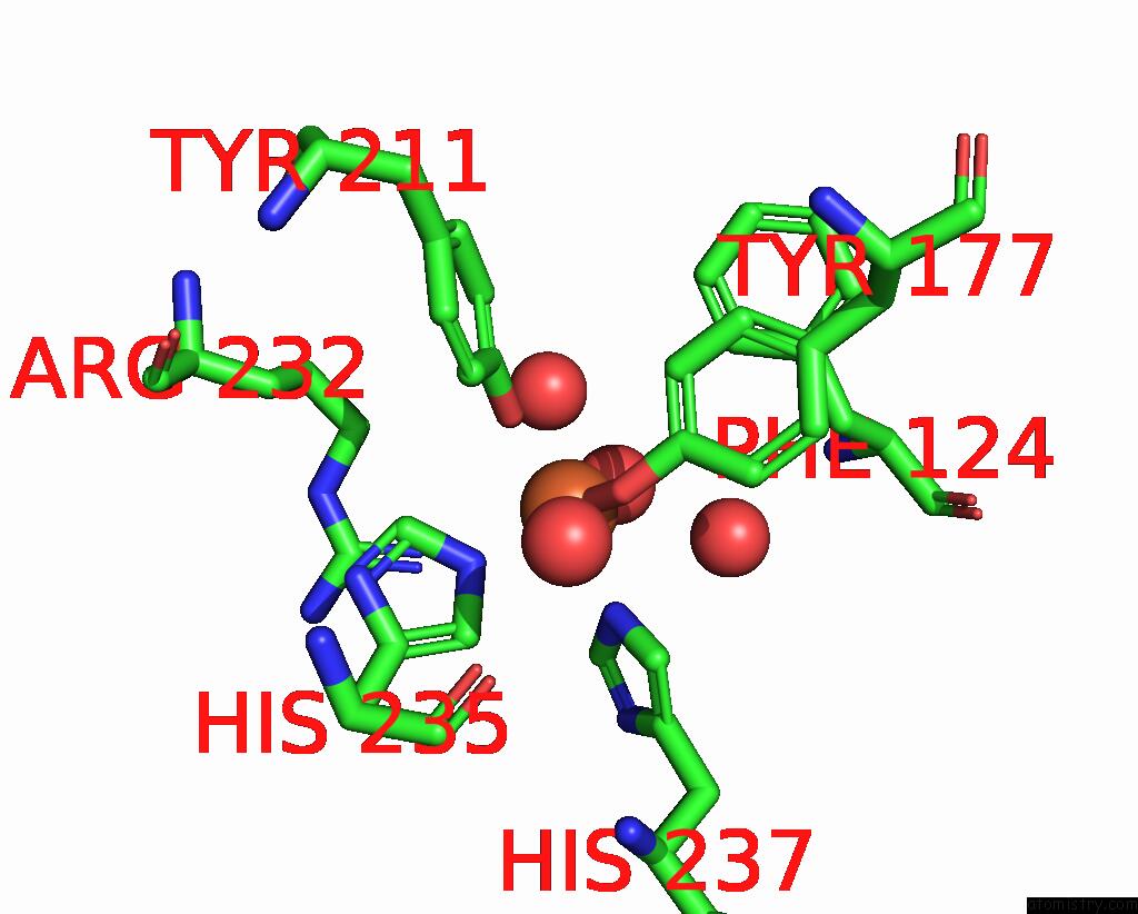
Mono view
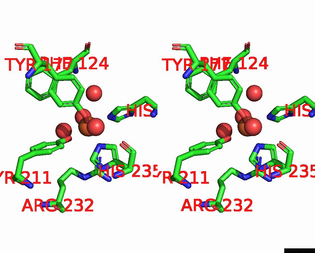
Stereo pair view
Mono view
Stereo pair view
A full contact list of Iron with other atoms in the Fe binding
site number 1 of Crystal Structure of Hydroxyquinol-1,2-Dioxygenase From Gelatoporia Subvermispora (GSHDX1) within 5.0Å range:
|
Iron binding site 2 out of 2 in 8r2v
Go back to
Iron binding site 2 out
of 2 in the Crystal Structure of Hydroxyquinol-1,2-Dioxygenase From Gelatoporia Subvermispora (GSHDX1)
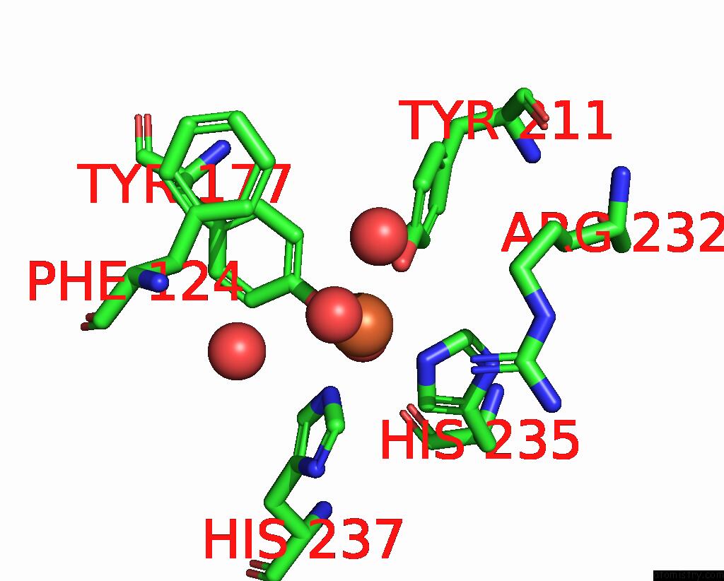
Mono view
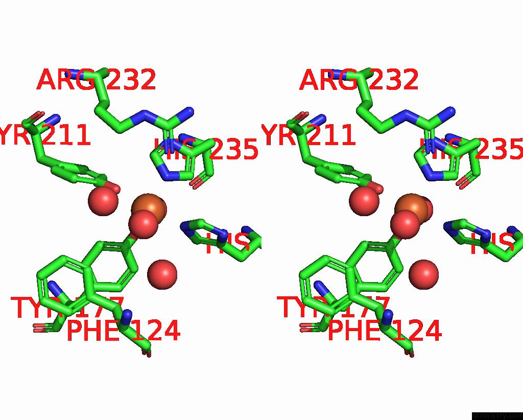
Stereo pair view
Mono view
Stereo pair view
A full contact list of Iron with other atoms in the Fe binding
site number 2 of Crystal Structure of Hydroxyquinol-1,2-Dioxygenase From Gelatoporia Subvermispora (GSHDX1) within 5.0Å range:
|
Reference:
A.Schwartz,
E.Kuatsjah,
M.Zahn,
D.Salvachua.
Biochemical and Structural Characterization Hydroquinone Catabolic Pathway From White-Rot Fungi To Be Published.
Page generated: Wed Nov 27 17:45:42 2024
Last articles
Zn in 9J0NZn in 9J0O
Zn in 9J0P
Zn in 9FJX
Zn in 9EKB
Zn in 9C0F
Zn in 9CAH
Zn in 9CH0
Zn in 9CH3
Zn in 9CH1