Iron »
PDB 8xnq-8z4q »
8yb0 »
Iron in PDB 8yb0: Xfel Crystal Structure of the Reduced Form of F393H P450BM3 with N- Enanthyl-L-Prolyl-L-Phenylalanine in Complex with Styrene
Enzymatic activity of Xfel Crystal Structure of the Reduced Form of F393H P450BM3 with N- Enanthyl-L-Prolyl-L-Phenylalanine in Complex with Styrene
All present enzymatic activity of Xfel Crystal Structure of the Reduced Form of F393H P450BM3 with N- Enanthyl-L-Prolyl-L-Phenylalanine in Complex with Styrene:
1.14.14.1; 1.6.2.4;
1.14.14.1; 1.6.2.4;
Protein crystallography data
The structure of Xfel Crystal Structure of the Reduced Form of F393H P450BM3 with N- Enanthyl-L-Prolyl-L-Phenylalanine in Complex with Styrene, PDB code: 8yb0
was solved by
S.Nagao,
W.Kuwano,
T.Tosha,
K.Yamashita,
J.K.Stanfield,
C.Kasai,
S.Ariyasu,
O.Shoji,
H.Sugimoto,
M.Kubo,
with X-Ray Crystallography technique. A brief refinement statistics is given in the table below:
| Resolution Low / High (Å) | 9.99 / 1.60 |
| Space group | P 21 21 21 |
| Cell size a, b, c (Å), α, β, γ (°) | 58.83, 128.43, 149.53, 90, 90, 90 |
| R / Rfree (%) | 17.5 / 20.5 |
Iron Binding Sites:
The binding sites of Iron atom in the Xfel Crystal Structure of the Reduced Form of F393H P450BM3 with N- Enanthyl-L-Prolyl-L-Phenylalanine in Complex with Styrene
(pdb code 8yb0). This binding sites where shown within
5.0 Angstroms radius around Iron atom.
In total 2 binding sites of Iron where determined in the Xfel Crystal Structure of the Reduced Form of F393H P450BM3 with N- Enanthyl-L-Prolyl-L-Phenylalanine in Complex with Styrene, PDB code: 8yb0:
Jump to Iron binding site number: 1; 2;
In total 2 binding sites of Iron where determined in the Xfel Crystal Structure of the Reduced Form of F393H P450BM3 with N- Enanthyl-L-Prolyl-L-Phenylalanine in Complex with Styrene, PDB code: 8yb0:
Jump to Iron binding site number: 1; 2;
Iron binding site 1 out of 2 in 8yb0
Go back to
Iron binding site 1 out
of 2 in the Xfel Crystal Structure of the Reduced Form of F393H P450BM3 with N- Enanthyl-L-Prolyl-L-Phenylalanine in Complex with Styrene
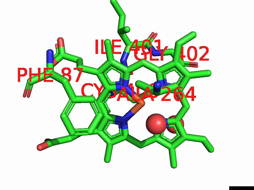
Mono view
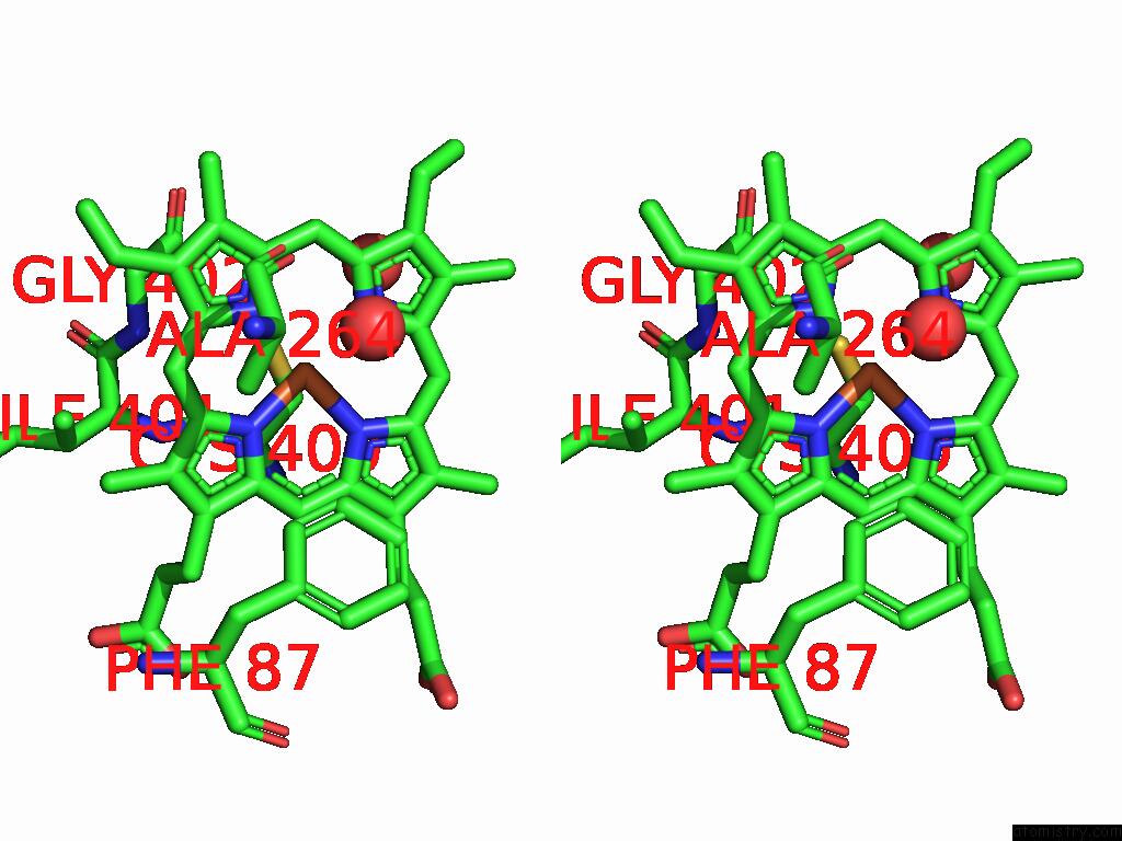
Stereo pair view
Mono view
Stereo pair view
A full contact list of Iron with other atoms in the Fe binding
site number 1 of Xfel Crystal Structure of the Reduced Form of F393H P450BM3 with N- Enanthyl-L-Prolyl-L-Phenylalanine in Complex with Styrene within 5.0Å range:
|
Iron binding site 2 out of 2 in 8yb0
Go back to
Iron binding site 2 out
of 2 in the Xfel Crystal Structure of the Reduced Form of F393H P450BM3 with N- Enanthyl-L-Prolyl-L-Phenylalanine in Complex with Styrene
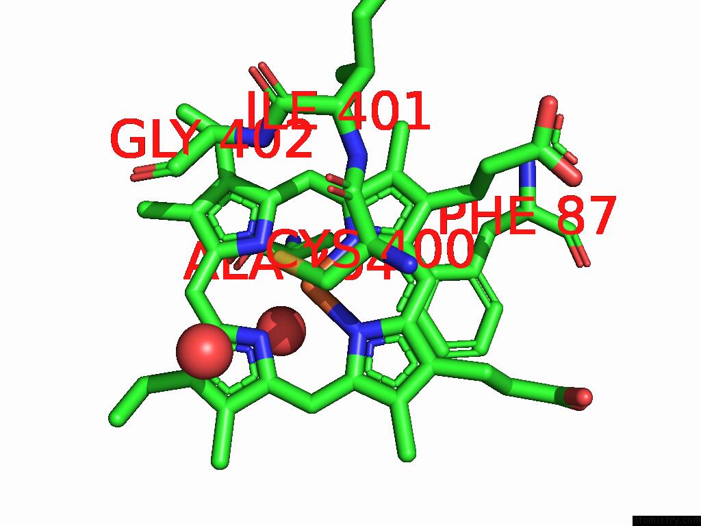
Mono view
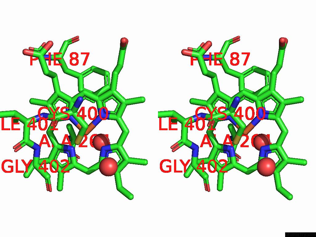
Stereo pair view
Mono view
Stereo pair view
A full contact list of Iron with other atoms in the Fe binding
site number 2 of Xfel Crystal Structure of the Reduced Form of F393H P450BM3 with N- Enanthyl-L-Prolyl-L-Phenylalanine in Complex with Styrene within 5.0Å range:
|
Reference:
S.Nagao,
W.Kuwano,
T.Tosha,
K.Yamashita,
J.K.Stanfield,
C.Kasai,
S.Ariyasu,
O.Shoji,
H.Sugimoto,
M.Kubo.
Xfel Crystallography Reveals Dynamic Control of Non-Natural Substrate Binding in Cytochrome P450BM3 Catalytic Cycle To Be Published.
Page generated: Tue Feb 25 09:56:46 2025
Last articles
Zn in 9MJ5Zn in 9HNW
Zn in 9G0L
Zn in 9FNE
Zn in 9DZN
Zn in 9E0I
Zn in 9D32
Zn in 9DAK
Zn in 8ZXC
Zn in 8ZUF