Iron »
PDB 9c8l-9etz »
9dfu »
Iron in PDB 9dfu: X-Ray Crystal Structure of the Second Viperin-Like Enzyme From T. Virens Variant F40H with Bound Ctp and Sam
Protein crystallography data
The structure of X-Ray Crystal Structure of the Second Viperin-Like Enzyme From T. Virens Variant F40H with Bound Ctp and Sam, PDB code: 9dfu
was solved by
J.C.Lachowicz,
J.B.Bonanno,
T.L.Grove,
with X-Ray Crystallography technique. A brief refinement statistics is given in the table below:
| Resolution Low / High (Å) | 19.66 / 2.30 |
| Space group | P 21 21 21 |
| Cell size a, b, c (Å), α, β, γ (°) | 37.137, 55.959, 152.81, 90, 90, 90 |
| R / Rfree (%) | 22.6 / 26 |
Other elements in 9dfu:
The structure of X-Ray Crystal Structure of the Second Viperin-Like Enzyme From T. Virens Variant F40H with Bound Ctp and Sam also contains other interesting chemical elements:
| Chlorine | (Cl) | 1 atom |
Iron Binding Sites:
The binding sites of Iron atom in the X-Ray Crystal Structure of the Second Viperin-Like Enzyme From T. Virens Variant F40H with Bound Ctp and Sam
(pdb code 9dfu). This binding sites where shown within
5.0 Angstroms radius around Iron atom.
In total 4 binding sites of Iron where determined in the X-Ray Crystal Structure of the Second Viperin-Like Enzyme From T. Virens Variant F40H with Bound Ctp and Sam, PDB code: 9dfu:
Jump to Iron binding site number: 1; 2; 3; 4;
In total 4 binding sites of Iron where determined in the X-Ray Crystal Structure of the Second Viperin-Like Enzyme From T. Virens Variant F40H with Bound Ctp and Sam, PDB code: 9dfu:
Jump to Iron binding site number: 1; 2; 3; 4;
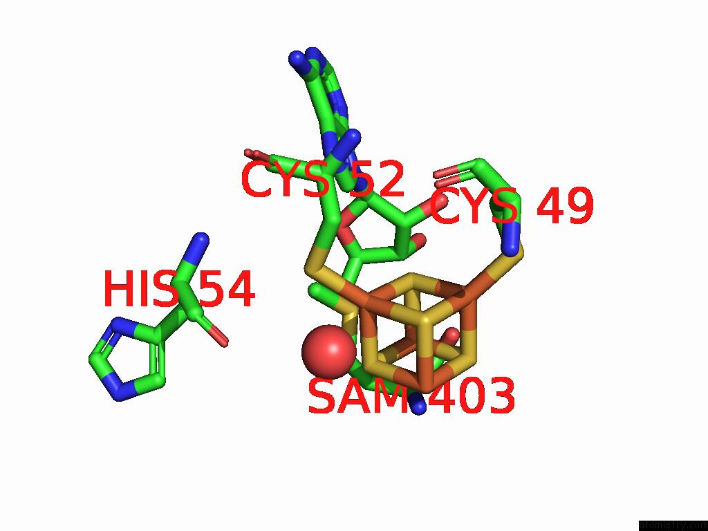
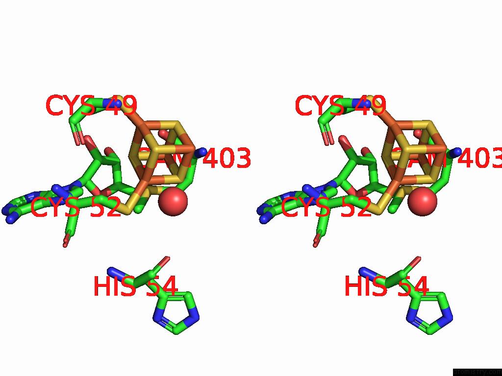
Iron binding site 1 out of 4 in 9dfu
Go back to
Iron binding site 1 out
of 4 in the X-Ray Crystal Structure of the Second Viperin-Like Enzyme From T. Virens Variant F40H with Bound Ctp and Sam
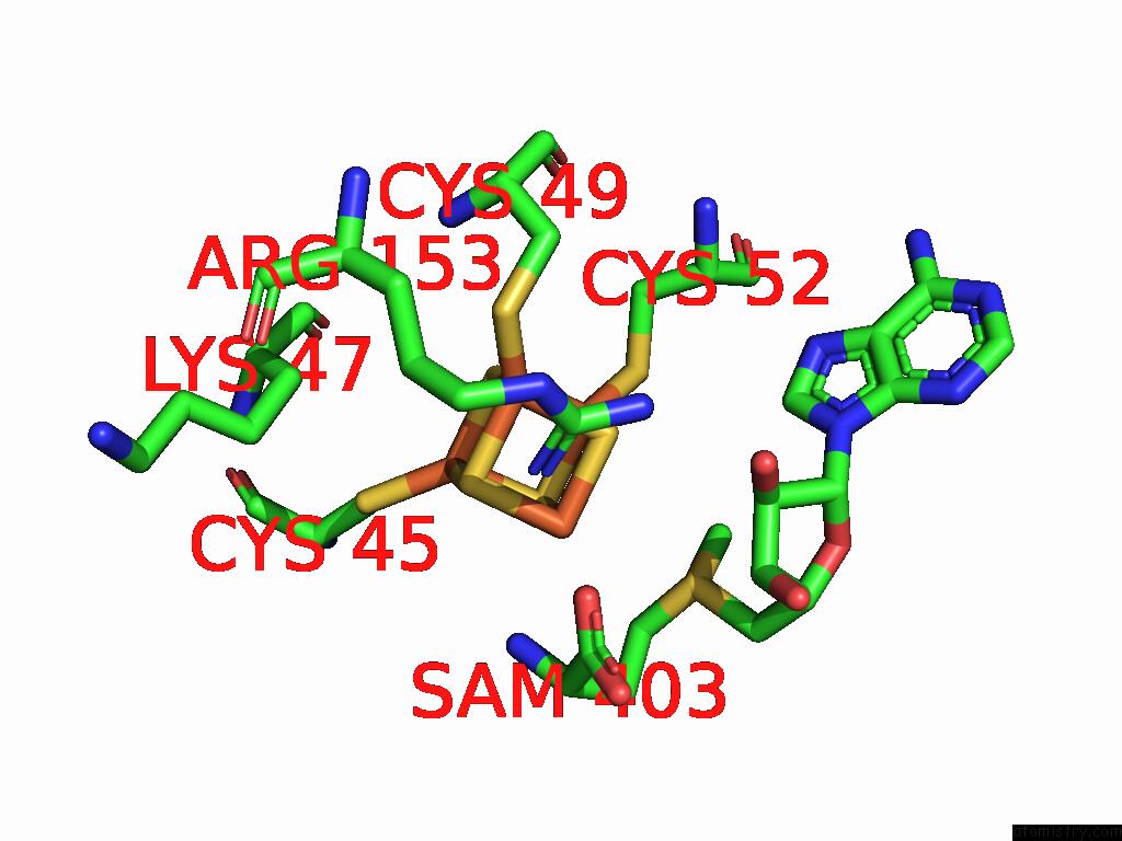
Mono view
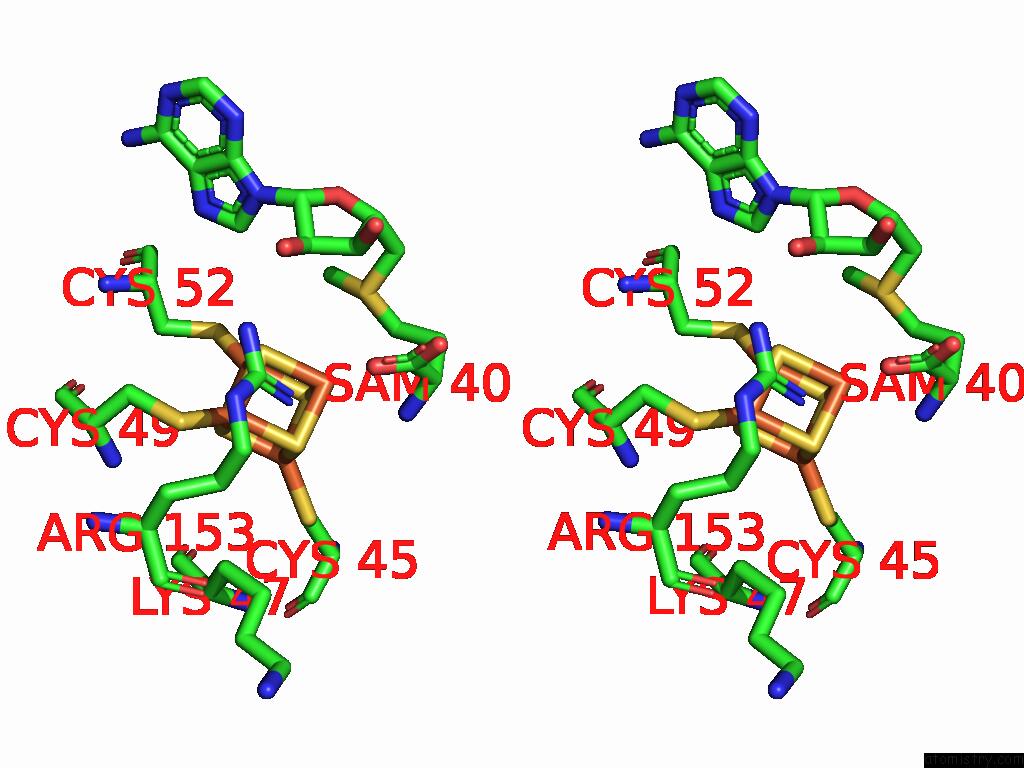
Stereo pair view
Mono view
Stereo pair view
A full contact list of Iron with other atoms in the Fe binding
site number 1 of X-Ray Crystal Structure of the Second Viperin-Like Enzyme From T. Virens Variant F40H with Bound Ctp and Sam within 5.0Å range:
|
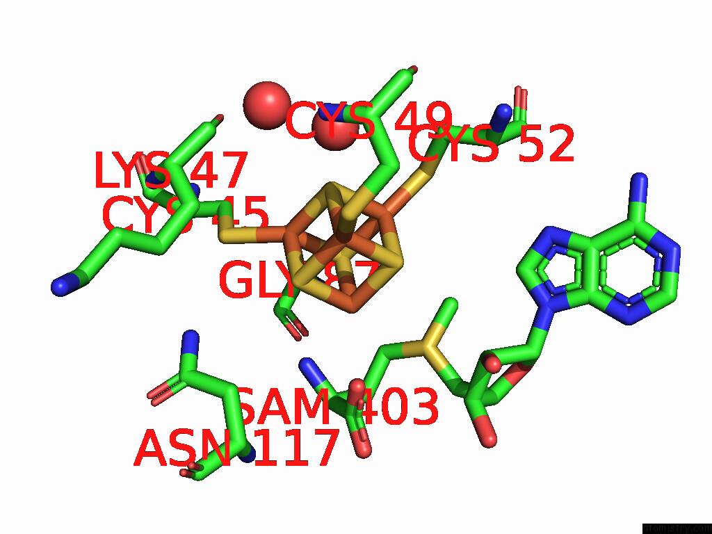
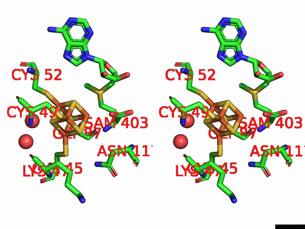
Iron binding site 2 out of 4 in 9dfu
Go back to
Iron binding site 2 out
of 4 in the X-Ray Crystal Structure of the Second Viperin-Like Enzyme From T. Virens Variant F40H with Bound Ctp and Sam
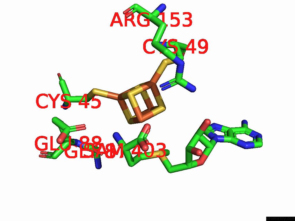
Mono view
Stereo pair view
Mono view
Stereo pair view
A full contact list of Iron with other atoms in the Fe binding
site number 2 of X-Ray Crystal Structure of the Second Viperin-Like Enzyme From T. Virens Variant F40H with Bound Ctp and Sam within 5.0Å range:
|
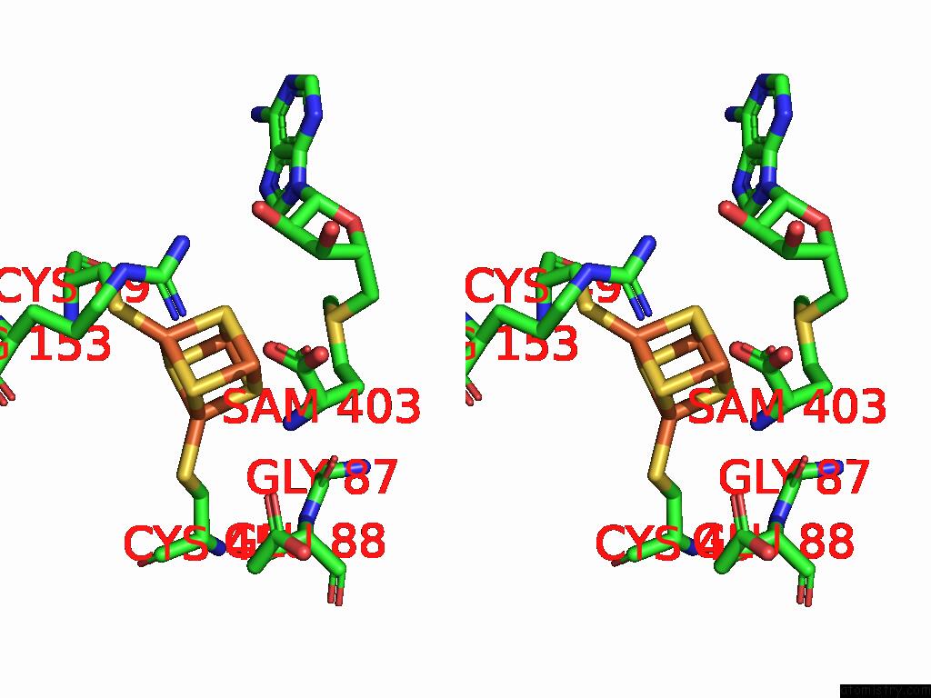
Iron binding site 3 out of 4 in 9dfu
Go back to
Iron binding site 3 out
of 4 in the X-Ray Crystal Structure of the Second Viperin-Like Enzyme From T. Virens Variant F40H with Bound Ctp and Sam
Mono view
Stereo pair view
Mono view
Stereo pair view
A full contact list of Iron with other atoms in the Fe binding
site number 3 of X-Ray Crystal Structure of the Second Viperin-Like Enzyme From T. Virens Variant F40H with Bound Ctp and Sam within 5.0Å range:
|
Iron binding site 4 out of 4 in 9dfu
Go back to
Iron binding site 4 out
of 4 in the X-Ray Crystal Structure of the Second Viperin-Like Enzyme From T. Virens Variant F40H with Bound Ctp and Sam
Mono view
Stereo pair view
Mono view
Stereo pair view
A full contact list of Iron with other atoms in the Fe binding
site number 4 of X-Ray Crystal Structure of the Second Viperin-Like Enzyme From T. Virens Variant F40H with Bound Ctp and Sam within 5.0Å range:
|
Reference:
J.C.Lachowicz,
S.Grudman,
J.B.Bonanno,
A.Fiser,
T.L.Grove.
Structural Insights From Active Site Variants and B-8 Loop Interactions in Viperin-Like Enzymes To Be Published.
Page generated: Tue Feb 25 09:58:26 2025
Last articles
Zn in 9MJ5Zn in 9HNW
Zn in 9G0L
Zn in 9FNE
Zn in 9DZN
Zn in 9E0I
Zn in 9D32
Zn in 9DAK
Zn in 8ZXC
Zn in 8ZUF