Iron »
PDB 9cqs-9e6x »
9dqq »
Iron in PDB 9dqq: Crystal Structure of Hrmj From Streptomyces Sp. AG109_G2-6 (Hrmj-Ssa) Complexed with Ferric Iron(III) and 2-Oxoglutarate
Protein crystallography data
The structure of Crystal Structure of Hrmj From Streptomyces Sp. AG109_G2-6 (Hrmj-Ssa) Complexed with Ferric Iron(III) and 2-Oxoglutarate, PDB code: 9dqq
was solved by
Y.-C.Zheng,
W.-C.Chang,
with X-Ray Crystallography technique. A brief refinement statistics is given in the table below:
| Resolution Low / High (Å) | 17.68 / 2.45 |
| Space group | C 1 2 1 |
| Cell size a, b, c (Å), α, β, γ (°) | 96.871, 43.616, 126.201, 90, 106.95, 90 |
| R / Rfree (%) | 20.7 / 28 |
Iron Binding Sites:
The binding sites of Iron atom in the Crystal Structure of Hrmj From Streptomyces Sp. AG109_G2-6 (Hrmj-Ssa) Complexed with Ferric Iron(III) and 2-Oxoglutarate
(pdb code 9dqq). This binding sites where shown within
5.0 Angstroms radius around Iron atom.
In total 3 binding sites of Iron where determined in the Crystal Structure of Hrmj From Streptomyces Sp. AG109_G2-6 (Hrmj-Ssa) Complexed with Ferric Iron(III) and 2-Oxoglutarate, PDB code: 9dqq:
Jump to Iron binding site number: 1; 2; 3;
In total 3 binding sites of Iron where determined in the Crystal Structure of Hrmj From Streptomyces Sp. AG109_G2-6 (Hrmj-Ssa) Complexed with Ferric Iron(III) and 2-Oxoglutarate, PDB code: 9dqq:
Jump to Iron binding site number: 1; 2; 3;
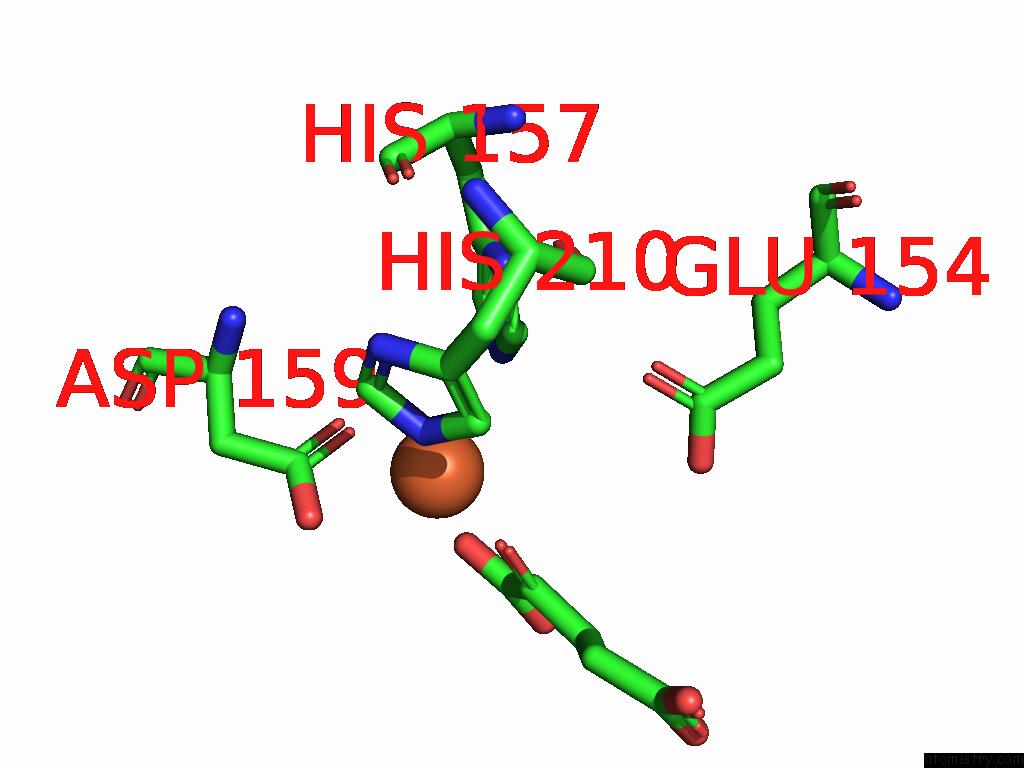
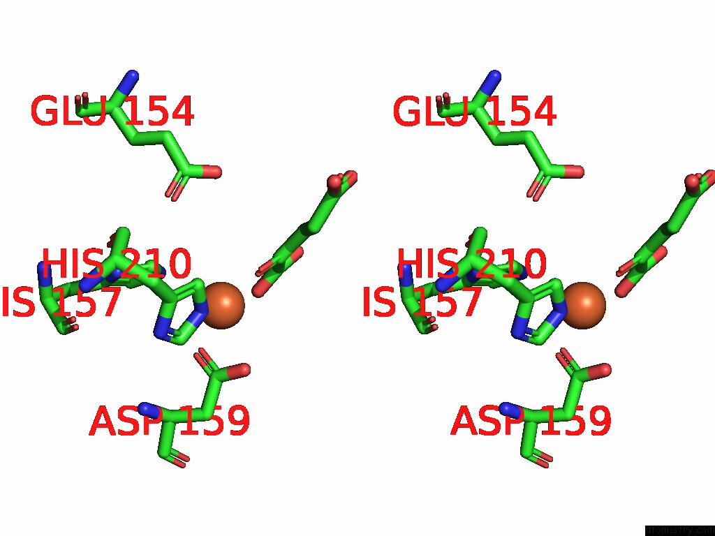
Iron binding site 1 out of 3 in 9dqq
Go back to
Iron binding site 1 out
of 3 in the Crystal Structure of Hrmj From Streptomyces Sp. AG109_G2-6 (Hrmj-Ssa) Complexed with Ferric Iron(III) and 2-Oxoglutarate
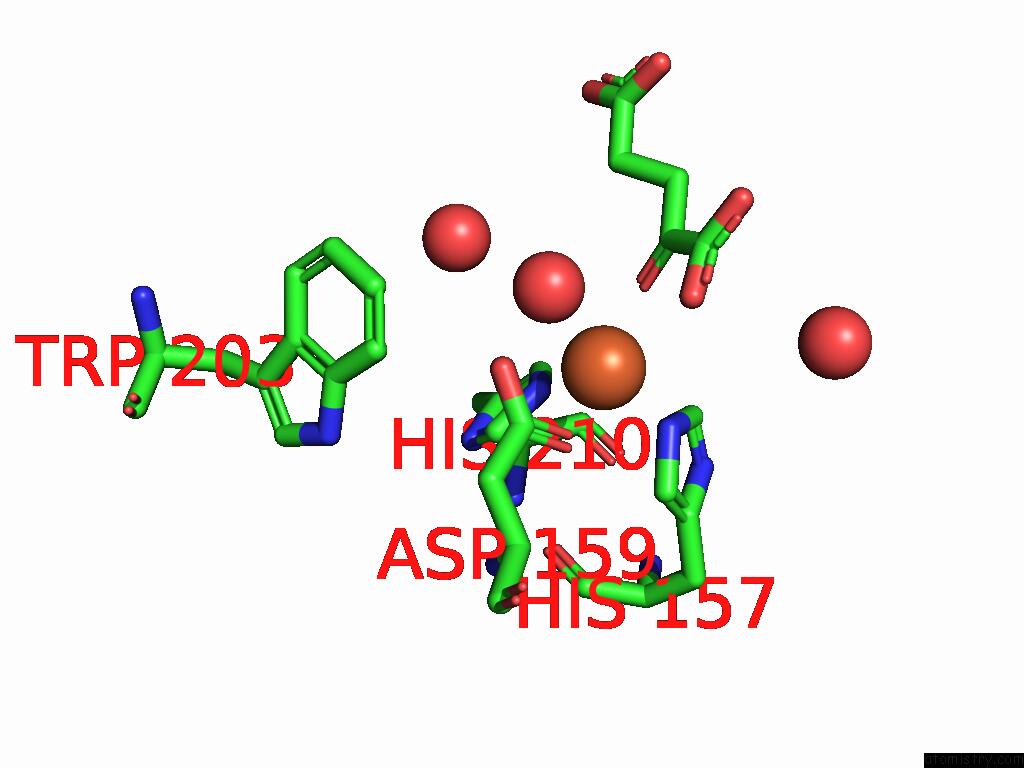
Mono view
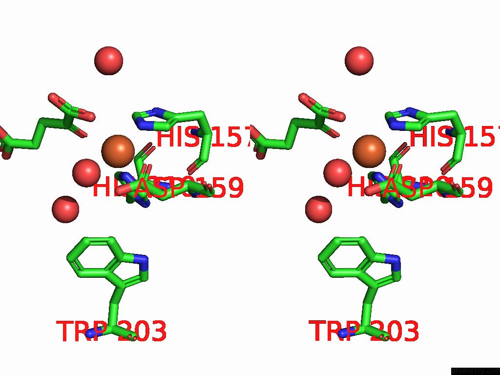
Stereo pair view
Mono view
Stereo pair view
A full contact list of Iron with other atoms in the Fe binding
site number 1 of Crystal Structure of Hrmj From Streptomyces Sp. AG109_G2-6 (Hrmj-Ssa) Complexed with Ferric Iron(III) and 2-Oxoglutarate within 5.0Å range:
|
Iron binding site 2 out of 3 in 9dqq
Go back to
Iron binding site 2 out
of 3 in the Crystal Structure of Hrmj From Streptomyces Sp. AG109_G2-6 (Hrmj-Ssa) Complexed with Ferric Iron(III) and 2-Oxoglutarate
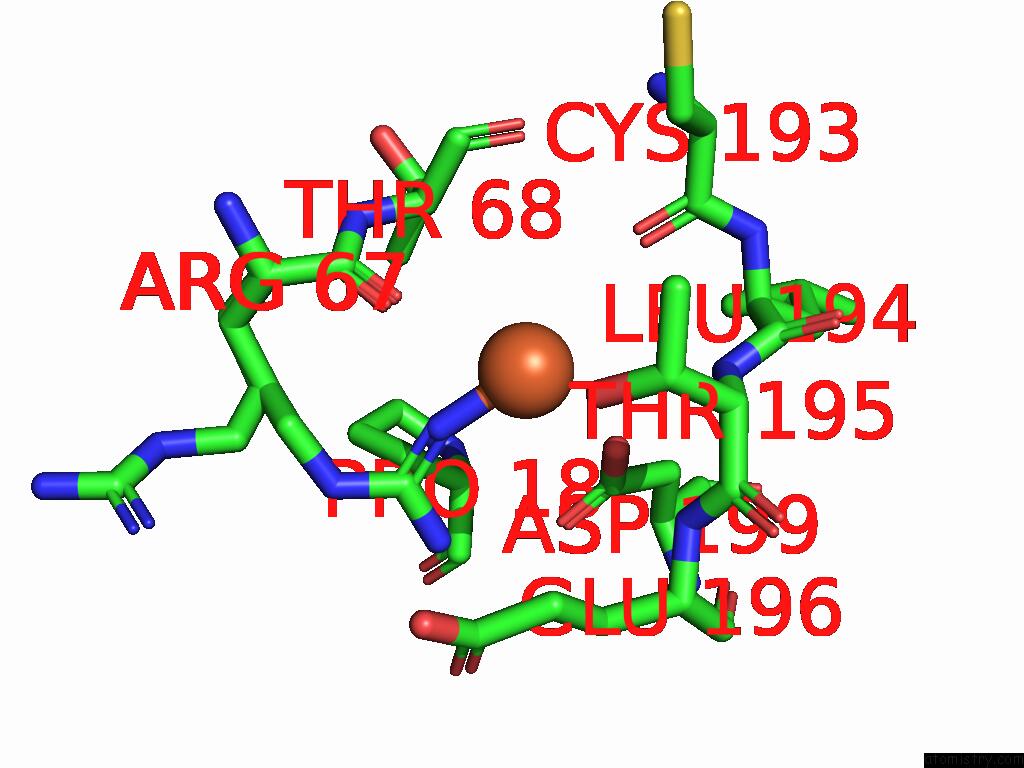
Mono view
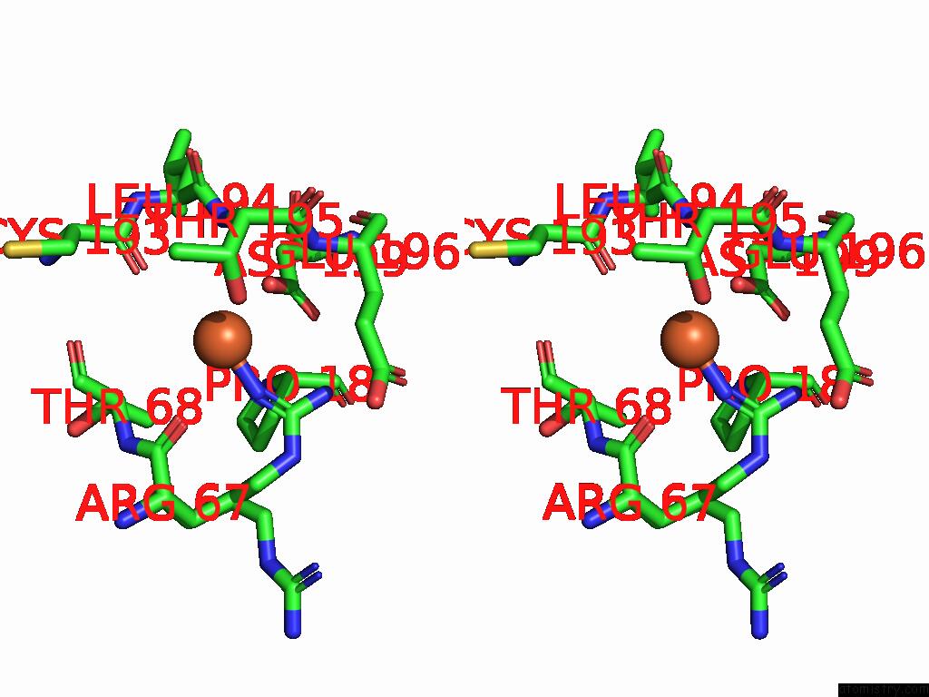
Stereo pair view
Mono view
Stereo pair view
A full contact list of Iron with other atoms in the Fe binding
site number 2 of Crystal Structure of Hrmj From Streptomyces Sp. AG109_G2-6 (Hrmj-Ssa) Complexed with Ferric Iron(III) and 2-Oxoglutarate within 5.0Å range:
|
Iron binding site 3 out of 3 in 9dqq
Go back to
Iron binding site 3 out
of 3 in the Crystal Structure of Hrmj From Streptomyces Sp. AG109_G2-6 (Hrmj-Ssa) Complexed with Ferric Iron(III) and 2-Oxoglutarate
Mono view
Stereo pair view
Mono view
Stereo pair view
A full contact list of Iron with other atoms in the Fe binding
site number 3 of Crystal Structure of Hrmj From Streptomyces Sp. AG109_G2-6 (Hrmj-Ssa) Complexed with Ferric Iron(III) and 2-Oxoglutarate within 5.0Å range:
|
Reference:
Y.C.Zheng,
X.Li,
L.Cha,
J.C.Paris,
C.Michael,
R.Ushimaru,
Y.Ogasawara,
I.Abe,
Y.Guo,
W.C.Chang.
Comparison of A Nonheme Iron Cyclopropanase with A Homologous Hydroxylase Reveals Mechanistic Features Associated with Distinct Reaction Outcomes. J.Am.Chem.Soc. 2025.
ISSN: ESSN 1520-5126
PubMed: 39901767
DOI: 10.1021/JACS.4C17741
Page generated: Fri Aug 8 03:49:47 2025
ISSN: ESSN 1520-5126
PubMed: 39901767
DOI: 10.1021/JACS.4C17741
Last articles
Fe in 9IIUFe in 9HNO
Fe in 9I2A
Fe in 9HNN
Fe in 9HXV
Fe in 9HNK
Fe in 9HNM
Fe in 9HNL
Fe in 9HIF
Fe in 9HI3