Iron »
PDB 9hfm-9iy1 »
9ixl »
Iron in PDB 9ixl: Crystal Structure of the CYP153A Double Mutant L354T/V456G From Marinobacter Aquaeolei
Protein crystallography data
The structure of Crystal Structure of the CYP153A Double Mutant L354T/V456G From Marinobacter Aquaeolei, PDB code: 9ixl
was solved by
M.M.Qin,
Y.P.Jiang,
Z.Q.Cong,
P.X.Zhao,
with X-Ray Crystallography technique. A brief refinement statistics is given in the table below:
| Resolution Low / High (Å) | 55.96 / 2.10 |
| Space group | C 2 2 21 |
| Cell size a, b, c (Å), α, β, γ (°) | 100.64, 102.02, 223.86, 90, 90, 90 |
| R / Rfree (%) | 19.6 / 21.9 |
Iron Binding Sites:
The binding sites of Iron atom in the Crystal Structure of the CYP153A Double Mutant L354T/V456G From Marinobacter Aquaeolei
(pdb code 9ixl). This binding sites where shown within
5.0 Angstroms radius around Iron atom.
In total 2 binding sites of Iron where determined in the Crystal Structure of the CYP153A Double Mutant L354T/V456G From Marinobacter Aquaeolei, PDB code: 9ixl:
Jump to Iron binding site number: 1; 2;
In total 2 binding sites of Iron where determined in the Crystal Structure of the CYP153A Double Mutant L354T/V456G From Marinobacter Aquaeolei, PDB code: 9ixl:
Jump to Iron binding site number: 1; 2;
Iron binding site 1 out of 2 in 9ixl
Go back to
Iron binding site 1 out
of 2 in the Crystal Structure of the CYP153A Double Mutant L354T/V456G From Marinobacter Aquaeolei
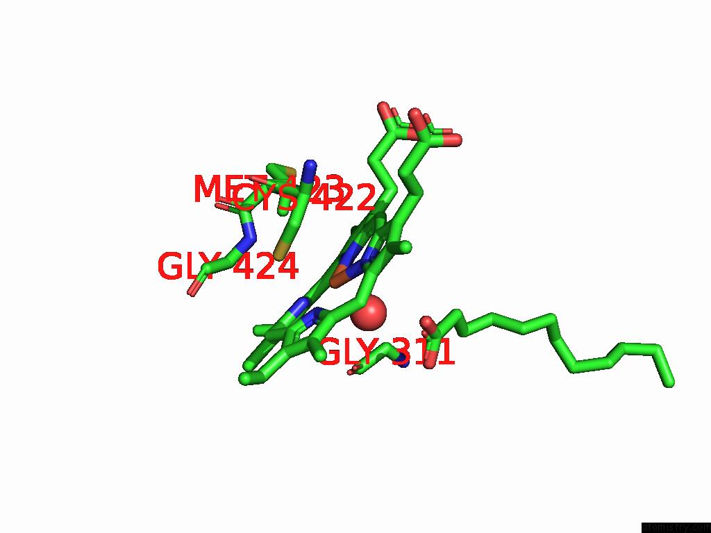
Mono view
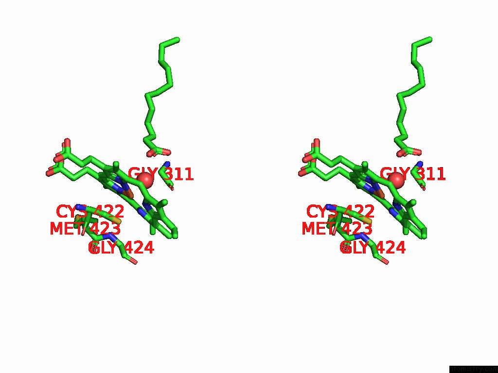
Stereo pair view
Mono view
Stereo pair view
A full contact list of Iron with other atoms in the Fe binding
site number 1 of Crystal Structure of the CYP153A Double Mutant L354T/V456G From Marinobacter Aquaeolei within 5.0Å range:
|
Iron binding site 2 out of 2 in 9ixl
Go back to
Iron binding site 2 out
of 2 in the Crystal Structure of the CYP153A Double Mutant L354T/V456G From Marinobacter Aquaeolei
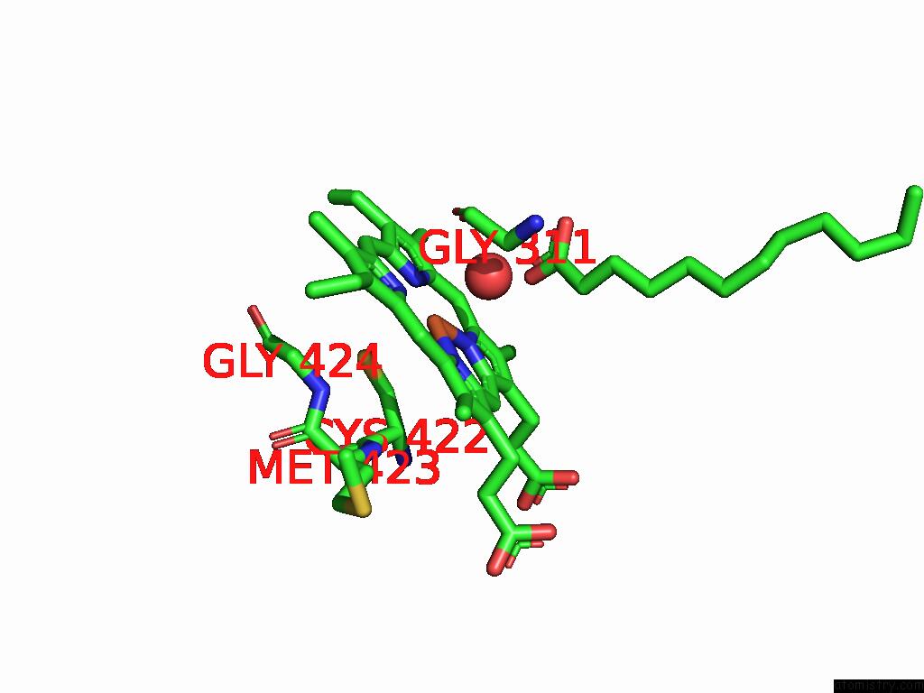
Mono view
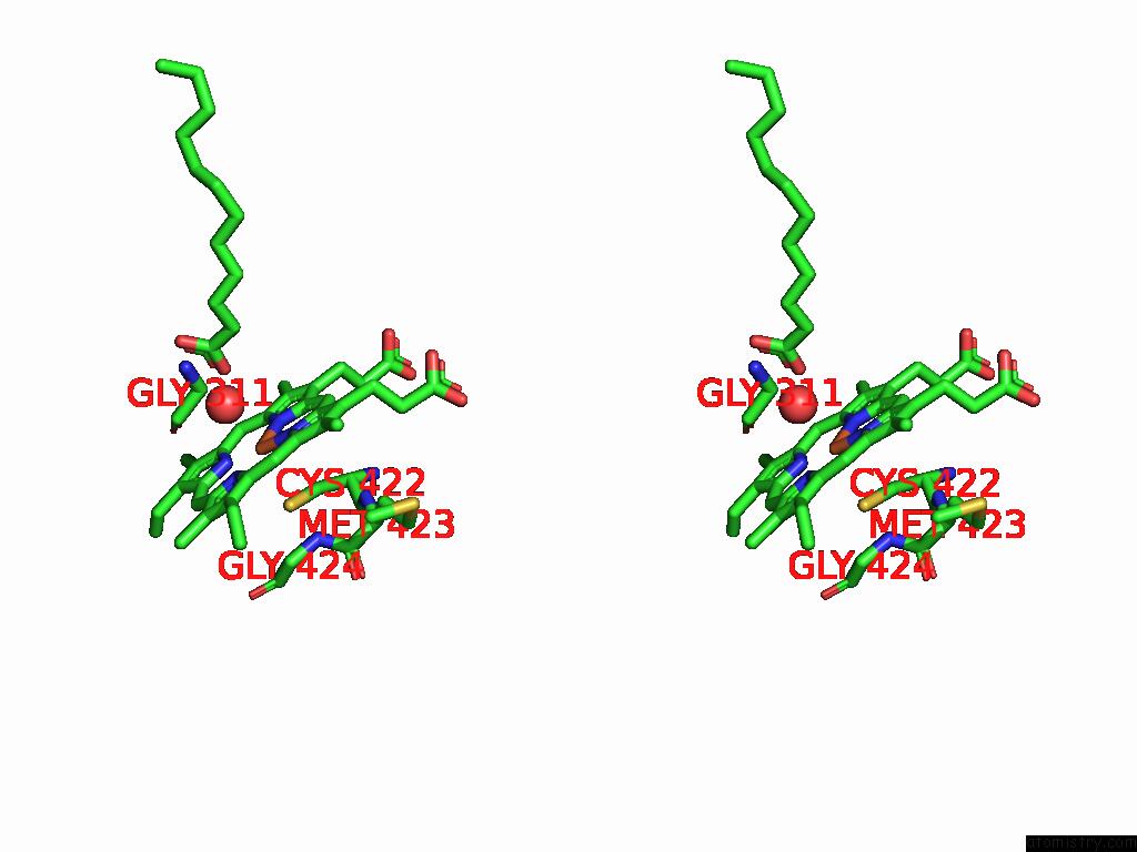
Stereo pair view
Mono view
Stereo pair view
A full contact list of Iron with other atoms in the Fe binding
site number 2 of Crystal Structure of the CYP153A Double Mutant L354T/V456G From Marinobacter Aquaeolei within 5.0Å range:
|
Reference:
M.M.Qin,
Y.P.Jiang,
Z.Q.Cong,
P.X.Zhao.
Structure of the CYP153A Double Mutant L354T/V456G From Marinobacter Aquaeolei at 2.10 Angstroms Resolution To Be Published.
Page generated: Sat Aug 23 03:20:00 2025
Last articles
Mn in 9LJUMn in 9LJW
Mn in 9LJS
Mn in 9LJR
Mn in 9LJT
Mn in 9LJV
Mg in 9UA2
Mg in 9R96
Mg in 9VM1
Mg in 9P01