Iron »
PDB 9ina-9kpp »
9jn8 »
Iron in PDB 9jn8: Crystal Structure of A Thioredoxin-Like Ferredoxin of Rhodobacter Capsulatus
Protein crystallography data
The structure of Crystal Structure of A Thioredoxin-Like Ferredoxin of Rhodobacter Capsulatus, PDB code: 9jn8
was solved by
Y.Shen,
L.Liu,
with X-Ray Crystallography technique. A brief refinement statistics is given in the table below:
| Resolution Low / High (Å) | 58.68 / 2.12 |
| Space group | P 41 2 2 |
| Cell size a, b, c (Å), α, β, γ (°) | 60.04, 60.04, 277.2, 90, 90, 90 |
| R / Rfree (%) | 20 / 24.1 |
Iron Binding Sites:
The binding sites of Iron atom in the Crystal Structure of A Thioredoxin-Like Ferredoxin of Rhodobacter Capsulatus
(pdb code 9jn8). This binding sites where shown within
5.0 Angstroms radius around Iron atom.
In total 6 binding sites of Iron where determined in the Crystal Structure of A Thioredoxin-Like Ferredoxin of Rhodobacter Capsulatus, PDB code: 9jn8:
Jump to Iron binding site number: 1; 2; 3; 4; 5; 6;
In total 6 binding sites of Iron where determined in the Crystal Structure of A Thioredoxin-Like Ferredoxin of Rhodobacter Capsulatus, PDB code: 9jn8:
Jump to Iron binding site number: 1; 2; 3; 4; 5; 6;
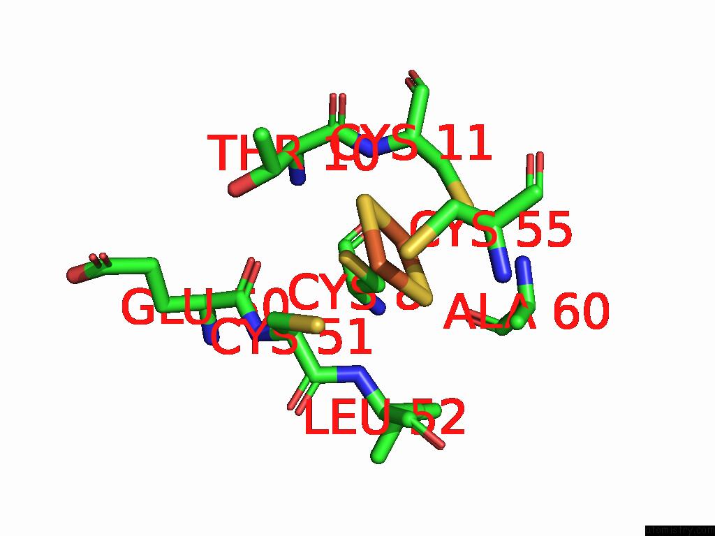
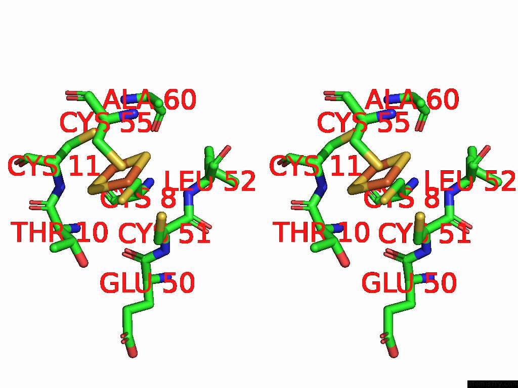
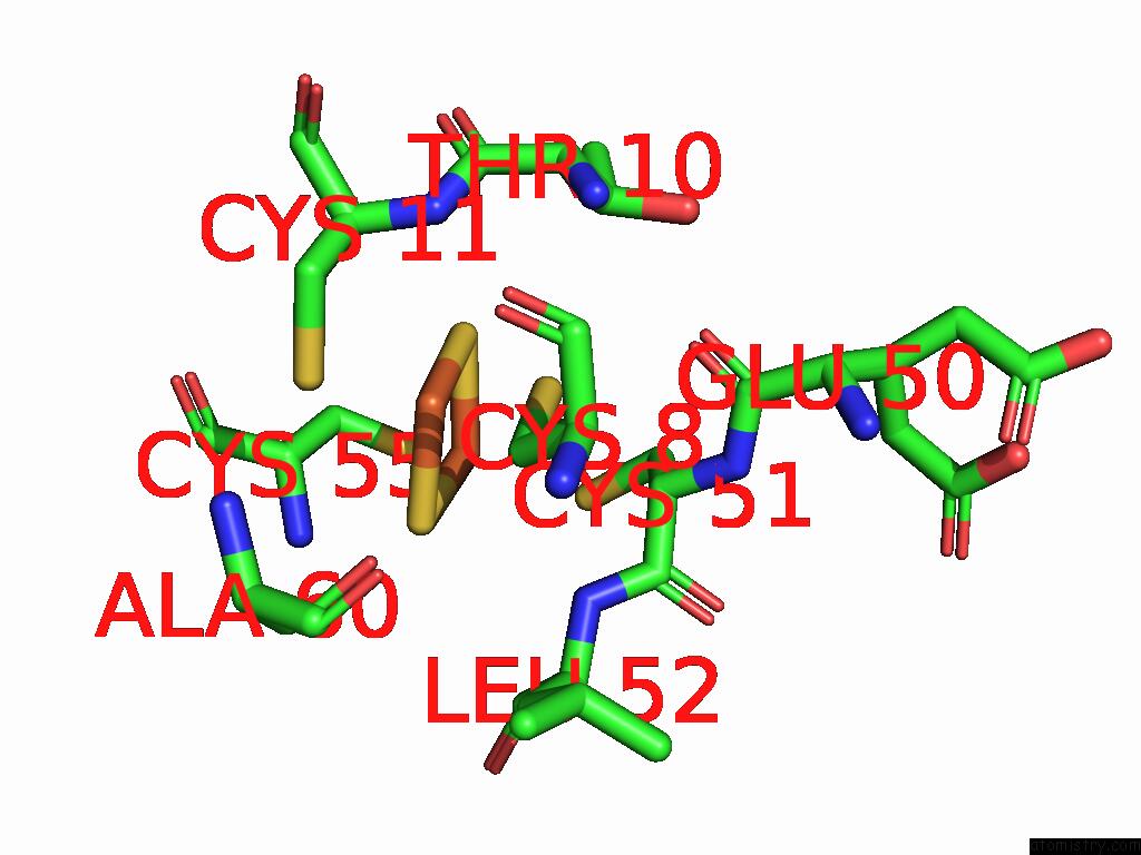
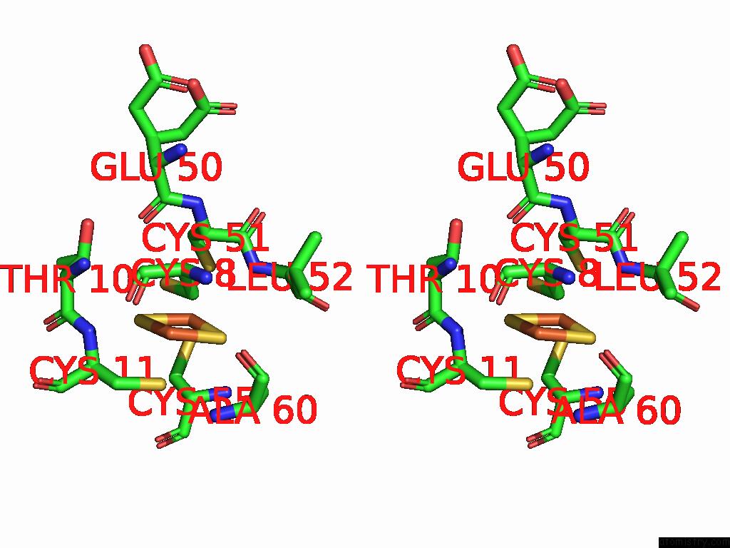
Iron binding site 1 out of 6 in 9jn8
Go back to
Iron binding site 1 out
of 6 in the Crystal Structure of A Thioredoxin-Like Ferredoxin of Rhodobacter Capsulatus
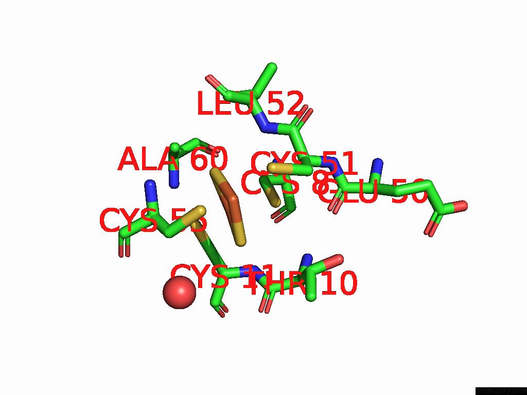
Mono view
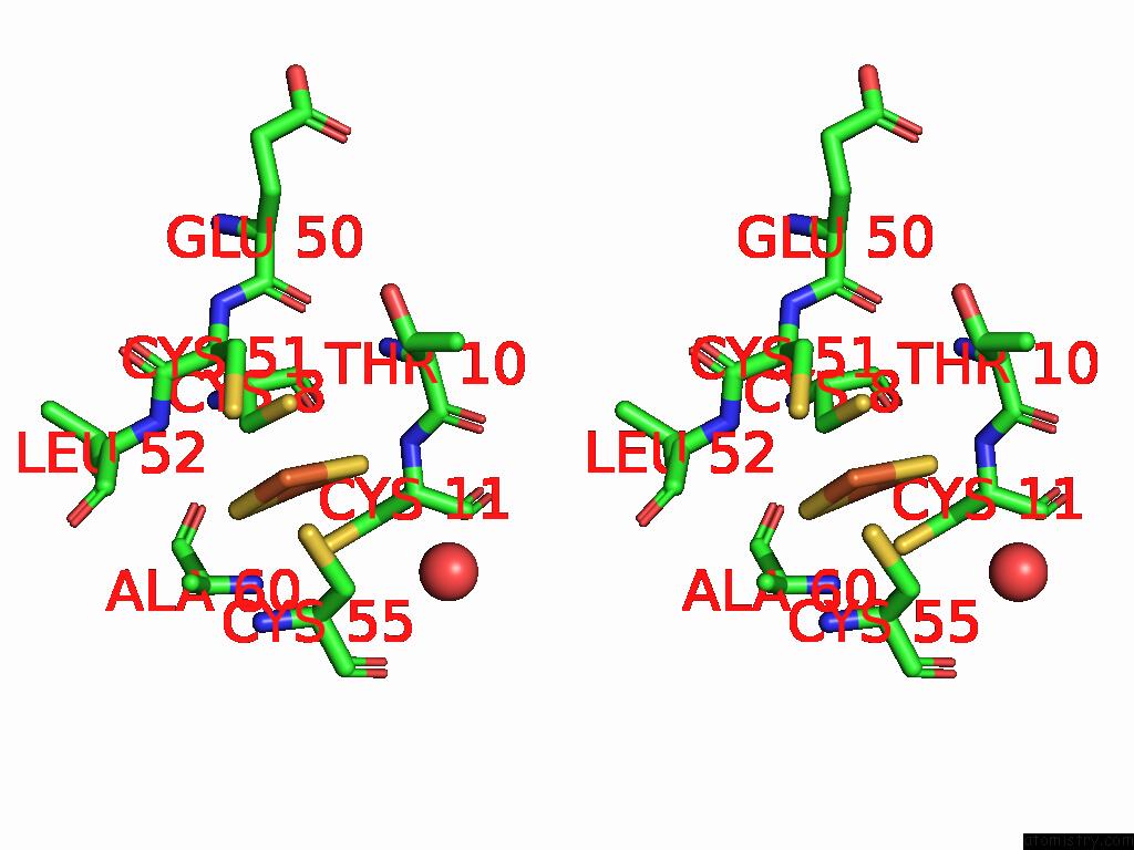
Stereo pair view
Mono view
Stereo pair view
A full contact list of Iron with other atoms in the Fe binding
site number 1 of Crystal Structure of A Thioredoxin-Like Ferredoxin of Rhodobacter Capsulatus within 5.0Å range:
|
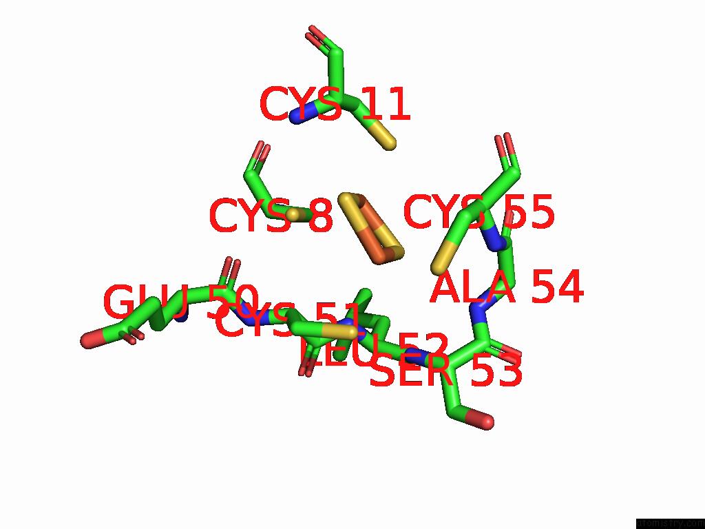
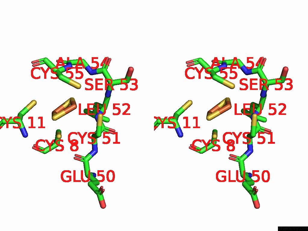
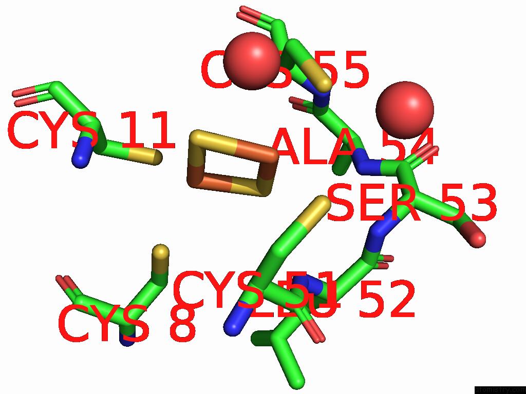
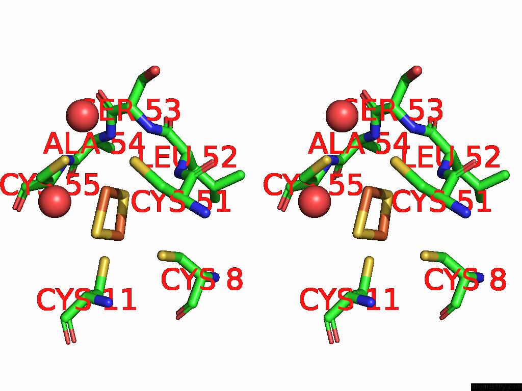
Iron binding site 2 out of 6 in 9jn8
Go back to
Iron binding site 2 out
of 6 in the Crystal Structure of A Thioredoxin-Like Ferredoxin of Rhodobacter Capsulatus
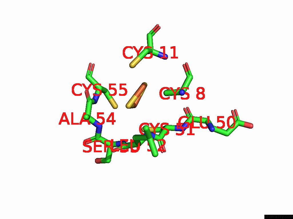
Mono view
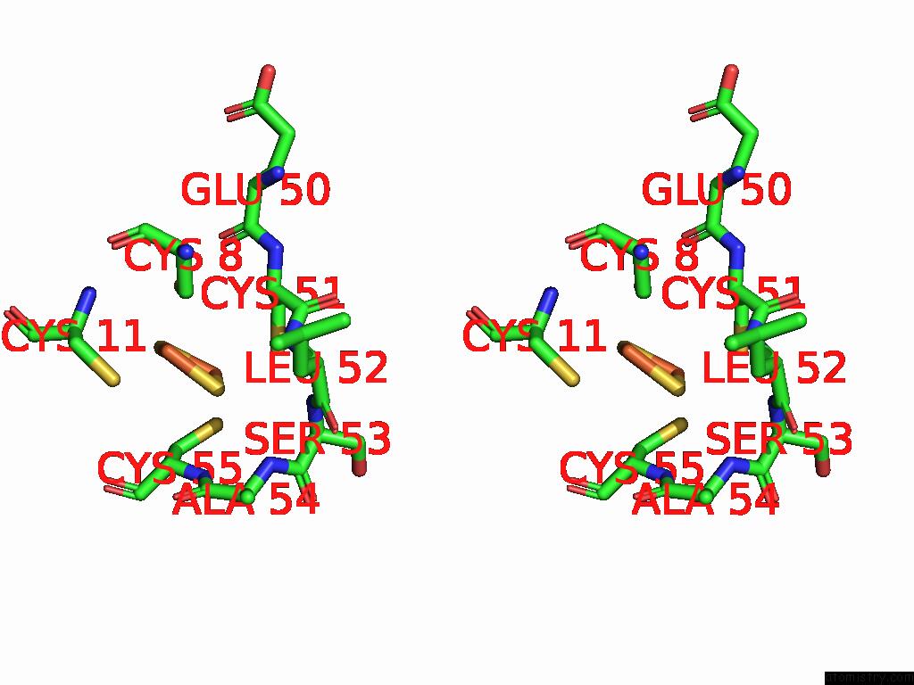
Stereo pair view
Mono view
Stereo pair view
A full contact list of Iron with other atoms in the Fe binding
site number 2 of Crystal Structure of A Thioredoxin-Like Ferredoxin of Rhodobacter Capsulatus within 5.0Å range:
|
Iron binding site 3 out of 6 in 9jn8
Go back to
Iron binding site 3 out
of 6 in the Crystal Structure of A Thioredoxin-Like Ferredoxin of Rhodobacter Capsulatus
Mono view
Stereo pair view
Mono view
Stereo pair view
A full contact list of Iron with other atoms in the Fe binding
site number 3 of Crystal Structure of A Thioredoxin-Like Ferredoxin of Rhodobacter Capsulatus within 5.0Å range:
|
Iron binding site 4 out of 6 in 9jn8
Go back to
Iron binding site 4 out
of 6 in the Crystal Structure of A Thioredoxin-Like Ferredoxin of Rhodobacter Capsulatus
Mono view
Stereo pair view
Mono view
Stereo pair view
A full contact list of Iron with other atoms in the Fe binding
site number 4 of Crystal Structure of A Thioredoxin-Like Ferredoxin of Rhodobacter Capsulatus within 5.0Å range:
|
Iron binding site 5 out of 6 in 9jn8
Go back to
Iron binding site 5 out
of 6 in the Crystal Structure of A Thioredoxin-Like Ferredoxin of Rhodobacter Capsulatus
Mono view
Stereo pair view
Mono view
Stereo pair view
A full contact list of Iron with other atoms in the Fe binding
site number 5 of Crystal Structure of A Thioredoxin-Like Ferredoxin of Rhodobacter Capsulatus within 5.0Å range:
|
Iron binding site 6 out of 6 in 9jn8
Go back to
Iron binding site 6 out
of 6 in the Crystal Structure of A Thioredoxin-Like Ferredoxin of Rhodobacter Capsulatus
Mono view
Stereo pair view
Mono view
Stereo pair view
A full contact list of Iron with other atoms in the Fe binding
site number 6 of Crystal Structure of A Thioredoxin-Like Ferredoxin of Rhodobacter Capsulatus within 5.0Å range:
|
Reference:
Y.H.Shen,
W.L.Cheng,
X.Wang,
H.E.Dai,
M.Wang,
L.Liu.
Crystal Structure of A Thioredoxin-Like Ferredoxin Encoded Within A Cobalamin Biosynthetic Operon of Rhodobacter Capsulatus. Protein J. 2025.
ISSN: ISSN 1572-3887
PubMed: 39924633
DOI: 10.1007/S10930-025-10254-Z
Page generated: Fri Aug 8 06:57:42 2025
ISSN: ISSN 1572-3887
PubMed: 39924633
DOI: 10.1007/S10930-025-10254-Z
Last articles
I in 1ORWI in 1OVL
I in 1OPM
I in 1OUQ
I in 1OJ5
I in 1OMK
I in 1NUO
I in 1NZB
I in 1O5B
I in 1NX3