Iron »
PDB 9g00-9nse »
9mt1 »
Iron in PDB 9mt1: 1.53 A Crystal Structure of Housefly Cytochrome C at pH 6.5
Protein crystallography data
The structure of 1.53 A Crystal Structure of Housefly Cytochrome C at pH 6.5, PDB code: 9mt1
was solved by
L.Zafreen,
L.-S.Huang,
P.Luemmen,
E.A.Berry,
with X-Ray Crystallography technique. A brief refinement statistics is given in the table below:
| Resolution Low / High (Å) | 29.19 / 1.41 |
| Space group | C 1 2 1 |
| Cell size a, b, c (Å), α, β, γ (°) | 58.222, 34.152, 105.071, 90, 105.04, 90 |
| R / Rfree (%) | 15.9 / 18.5 |
Other elements in 9mt1:
The structure of 1.53 A Crystal Structure of Housefly Cytochrome C at pH 6.5 also contains other interesting chemical elements:
| Sodium | (Na) | 1 atom |
Iron Binding Sites:
The binding sites of Iron atom in the 1.53 A Crystal Structure of Housefly Cytochrome C at pH 6.5
(pdb code 9mt1). This binding sites where shown within
5.0 Angstroms radius around Iron atom.
In total 2 binding sites of Iron where determined in the 1.53 A Crystal Structure of Housefly Cytochrome C at pH 6.5, PDB code: 9mt1:
Jump to Iron binding site number: 1; 2;
In total 2 binding sites of Iron where determined in the 1.53 A Crystal Structure of Housefly Cytochrome C at pH 6.5, PDB code: 9mt1:
Jump to Iron binding site number: 1; 2;
Iron binding site 1 out of 2 in 9mt1
Go back to
Iron binding site 1 out
of 2 in the 1.53 A Crystal Structure of Housefly Cytochrome C at pH 6.5
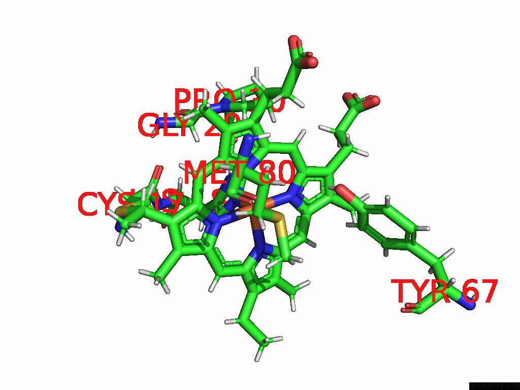
Mono view
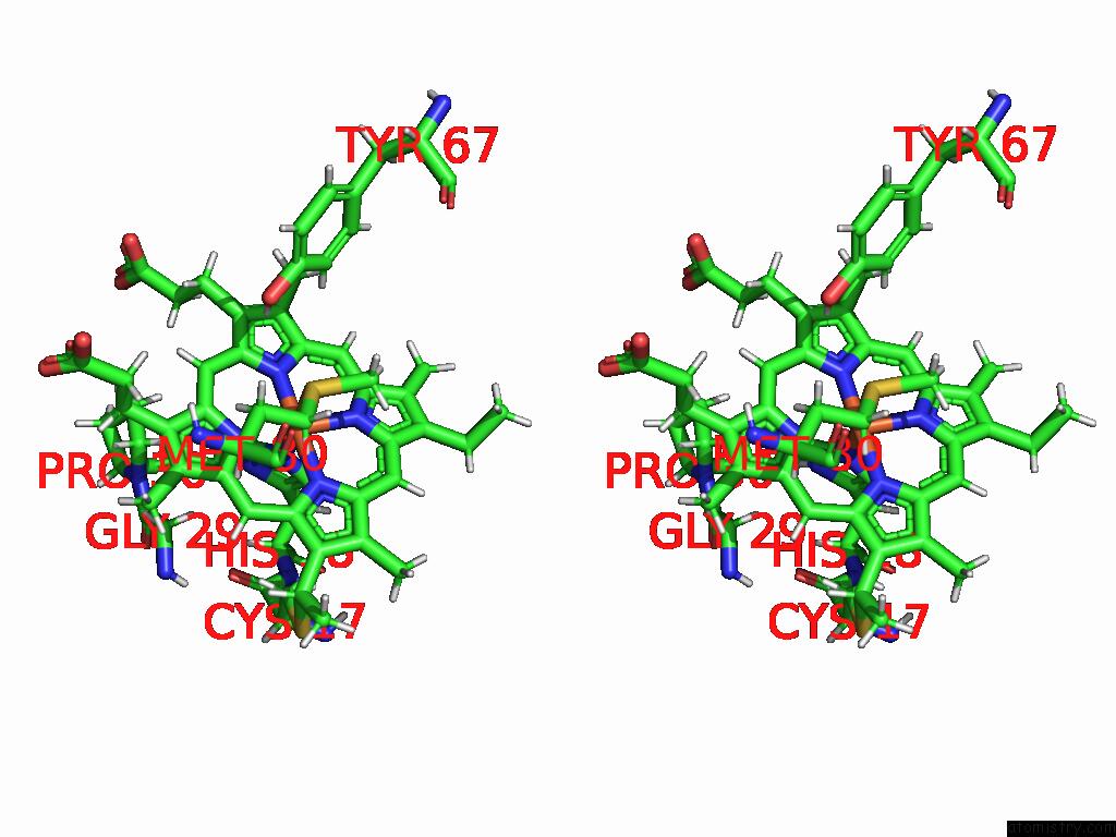
Stereo pair view
Mono view
Stereo pair view
A full contact list of Iron with other atoms in the Fe binding
site number 1 of 1.53 A Crystal Structure of Housefly Cytochrome C at pH 6.5 within 5.0Å range:
|
Iron binding site 2 out of 2 in 9mt1
Go back to
Iron binding site 2 out
of 2 in the 1.53 A Crystal Structure of Housefly Cytochrome C at pH 6.5
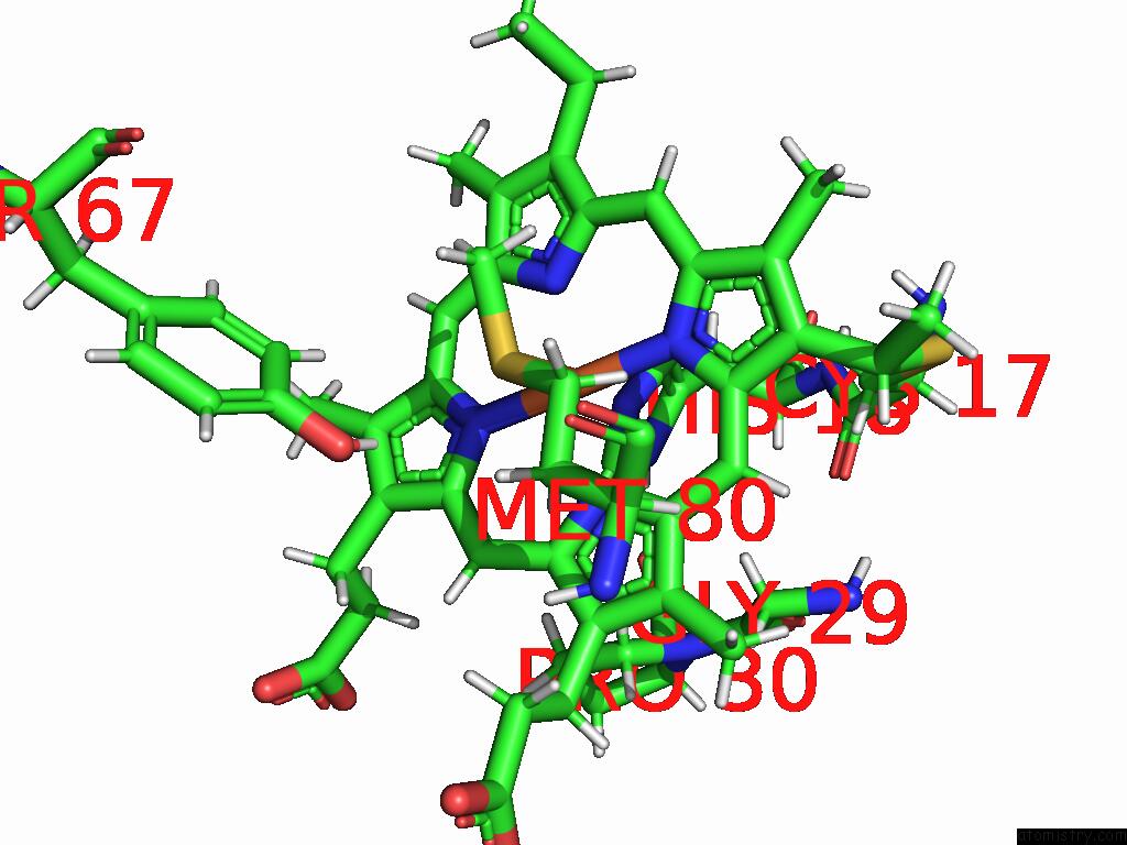
Mono view
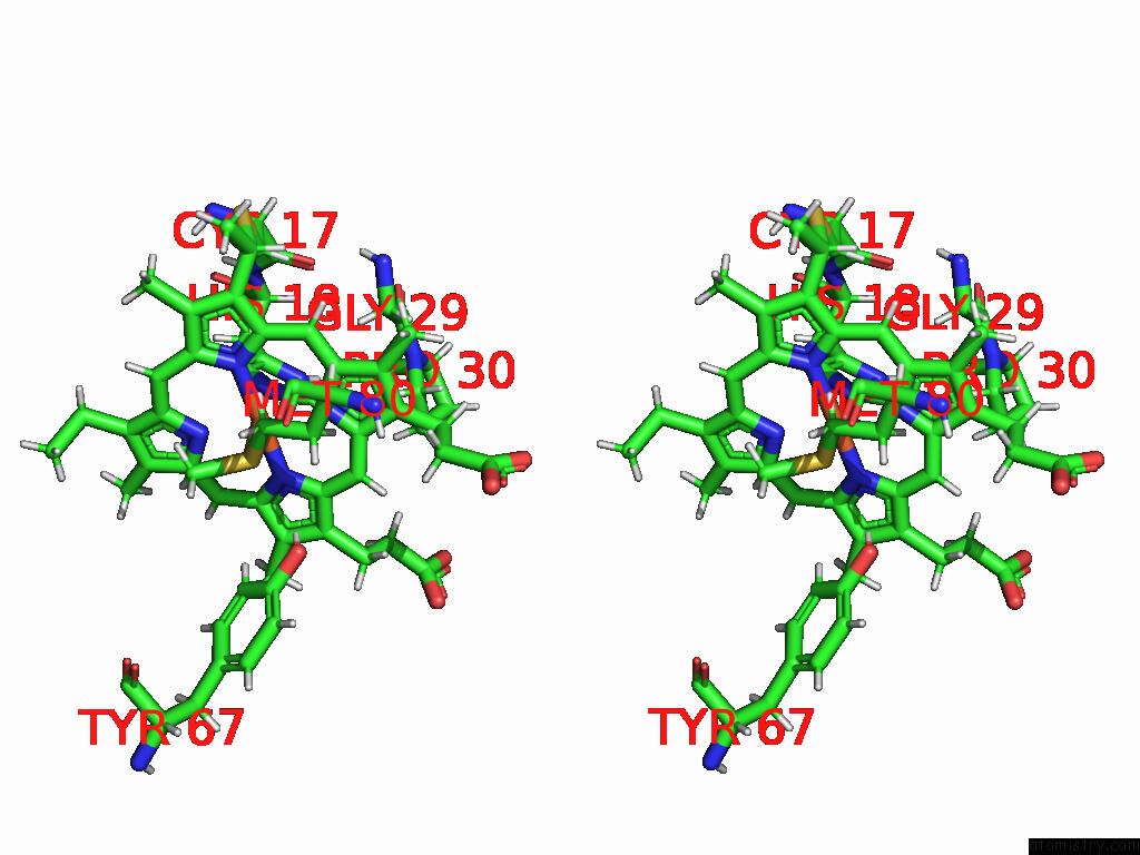
Stereo pair view
Mono view
Stereo pair view
A full contact list of Iron with other atoms in the Fe binding
site number 2 of 1.53 A Crystal Structure of Housefly Cytochrome C at pH 6.5 within 5.0Å range:
|
Reference:
L.Zafreen,
L.-S.Huang,
P.Luemmen,
E.A.Berry.
Crystal Structure of Insect Cytochrome C at Different pH Values. To Be Published.
Page generated: Tue Feb 25 10:02:49 2025
Last articles
Zn in 9MJ5Zn in 9HNW
Zn in 9G0L
Zn in 9FNE
Zn in 9DZN
Zn in 9E0I
Zn in 9D32
Zn in 9DAK
Zn in 8ZXC
Zn in 8ZUF