Iron »
PDB 2grz-2hk6 »
2h4i »
Iron in PDB 2h4i: Crystal Structure of the Complex of Proteolytically Produced C- Terminal Half of Bovine Lactoferrin with Lactose at 2.55 A Resolution
Protein crystallography data
The structure of Crystal Structure of the Complex of Proteolytically Produced C- Terminal Half of Bovine Lactoferrin with Lactose at 2.55 A Resolution, PDB code: 2h4i
was solved by
R.Mir,
R.Prem Kumar,
M.Sinha,
N.Singh,
P.Kaur,
S.Sharma,
T.P.Singh,
with X-Ray Crystallography technique. A brief refinement statistics is given in the table below:
| Resolution Low / High (Å) | 20.00 / 2.55 |
| Space group | P 1 21 1 |
| Cell size a, b, c (Å), α, β, γ (°) | 63.441, 50.417, 65.890, 90.00, 107.84, 90.00 |
| R / Rfree (%) | 18.8 / 21.6 |
Other elements in 2h4i:
The structure of Crystal Structure of the Complex of Proteolytically Produced C- Terminal Half of Bovine Lactoferrin with Lactose at 2.55 A Resolution also contains other interesting chemical elements:
| Zinc | (Zn) | 2 atoms |
Iron Binding Sites:
The binding sites of Iron atom in the Crystal Structure of the Complex of Proteolytically Produced C- Terminal Half of Bovine Lactoferrin with Lactose at 2.55 A Resolution
(pdb code 2h4i). This binding sites where shown within
5.0 Angstroms radius around Iron atom.
In total only one binding site of Iron was determined in the Crystal Structure of the Complex of Proteolytically Produced C- Terminal Half of Bovine Lactoferrin with Lactose at 2.55 A Resolution, PDB code: 2h4i:
In total only one binding site of Iron was determined in the Crystal Structure of the Complex of Proteolytically Produced C- Terminal Half of Bovine Lactoferrin with Lactose at 2.55 A Resolution, PDB code: 2h4i:
Iron binding site 1 out of 1 in 2h4i
Go back to
Iron binding site 1 out
of 1 in the Crystal Structure of the Complex of Proteolytically Produced C- Terminal Half of Bovine Lactoferrin with Lactose at 2.55 A Resolution
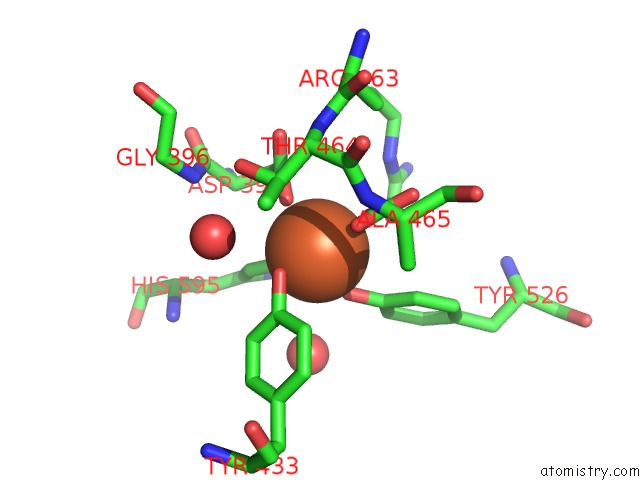
Mono view
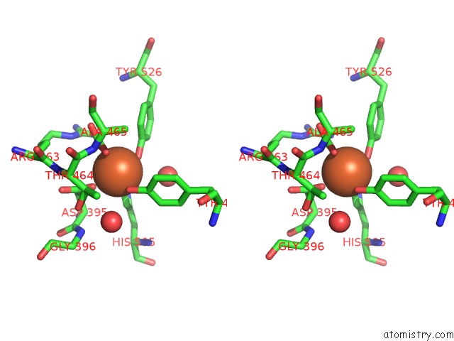
Stereo pair view
Mono view
Stereo pair view
A full contact list of Iron with other atoms in the Fe binding
site number 1 of Crystal Structure of the Complex of Proteolytically Produced C- Terminal Half of Bovine Lactoferrin with Lactose at 2.55 A Resolution within 5.0Å range:
|
Reference:
R.Mir,
R.Prem Kumar,
M.Sinha,
N.Singh,
P.Kaur,
S.Sharma,
T.P.Singh.
Crystal Structure of the Complex of Proteolytically Produced C-Terminal Half of Bovine Lactoferrin with Lactose at 2.55 A Resolution To Be Published.
Page generated: Thu Jul 17 01:55:55 2025
Last articles
Fe in 8CM6Fe in 8CM4
Fe in 8CH9
Fe in 8CKS
Fe in 8CKN
Fe in 8CK9
Fe in 8CJO
Fe in 8CJM
Fe in 8CJN
Fe in 8CJL