Iron »
PDB 2pgh-2q9u »
2pgh »
Iron in PDB 2pgh: Structure Determination of Aquomet Porcine Hemoglobin at 2.8 Angstrom Resolution
Protein crystallography data
The structure of Structure Determination of Aquomet Porcine Hemoglobin at 2.8 Angstrom Resolution, PDB code: 2pgh
was solved by
D.S.Katz,
S.P.White,
W.Huang,
R.Kumar,
D.W.Christianson,
with X-Ray Crystallography technique. A brief refinement statistics is given in the table below:
| Resolution Low / High (Å) | 6.50 / 2.80 |
| Space group | P 21 21 21 |
| Cell size a, b, c (Å), α, β, γ (°) | 69.600, 72.800, 115.800, 90.00, 90.00, 90.00 |
| R / Rfree (%) | 15.4 / n/a |
Iron Binding Sites:
The binding sites of Iron atom in the Structure Determination of Aquomet Porcine Hemoglobin at 2.8 Angstrom Resolution
(pdb code 2pgh). This binding sites where shown within
5.0 Angstroms radius around Iron atom.
In total 4 binding sites of Iron where determined in the Structure Determination of Aquomet Porcine Hemoglobin at 2.8 Angstrom Resolution, PDB code: 2pgh:
Jump to Iron binding site number: 1; 2; 3; 4;
In total 4 binding sites of Iron where determined in the Structure Determination of Aquomet Porcine Hemoglobin at 2.8 Angstrom Resolution, PDB code: 2pgh:
Jump to Iron binding site number: 1; 2; 3; 4;
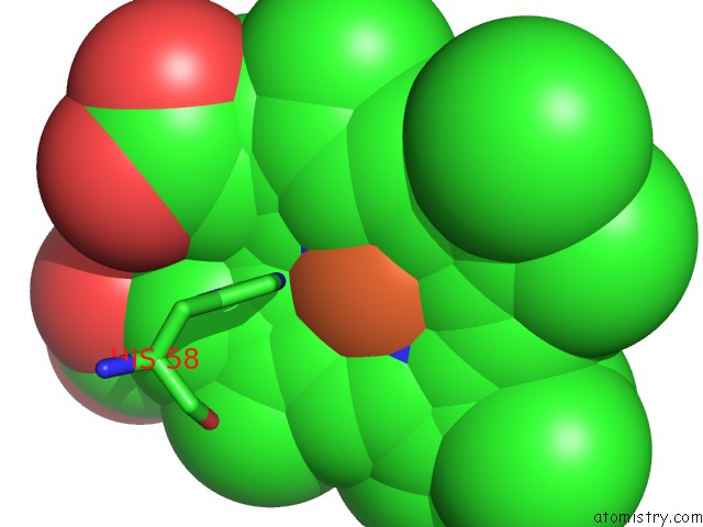
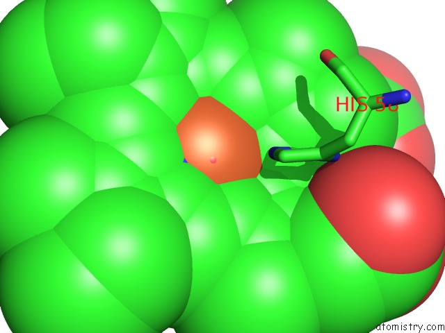
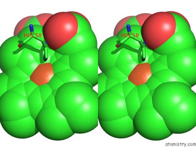
Iron binding site 1 out of 4 in 2pgh
Go back to
Iron binding site 1 out
of 4 in the Structure Determination of Aquomet Porcine Hemoglobin at 2.8 Angstrom Resolution
Mono view
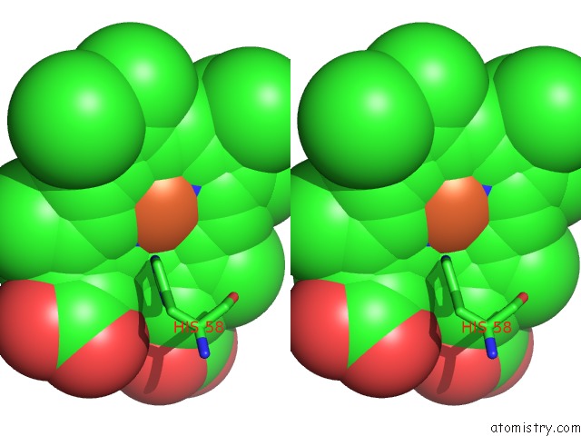
Stereo pair view
Mono view
Stereo pair view
A full contact list of Iron with other atoms in the Fe binding
site number 1 of Structure Determination of Aquomet Porcine Hemoglobin at 2.8 Angstrom Resolution within 5.0Å range:
|
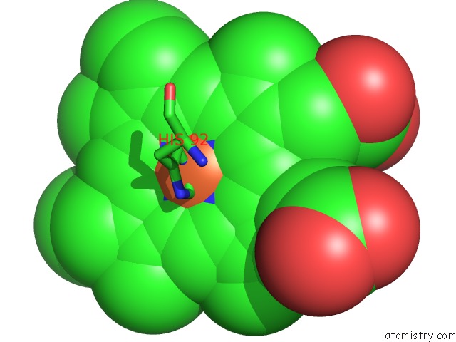
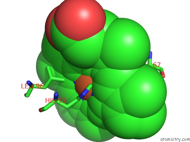
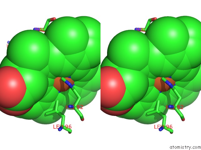
Iron binding site 2 out of 4 in 2pgh
Go back to
Iron binding site 2 out
of 4 in the Structure Determination of Aquomet Porcine Hemoglobin at 2.8 Angstrom Resolution
Mono view
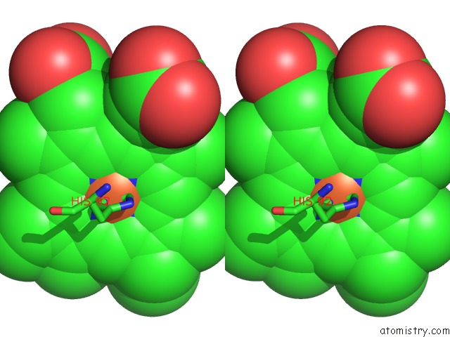
Stereo pair view
Mono view
Stereo pair view
A full contact list of Iron with other atoms in the Fe binding
site number 2 of Structure Determination of Aquomet Porcine Hemoglobin at 2.8 Angstrom Resolution within 5.0Å range:
|
Iron binding site 3 out of 4 in 2pgh
Go back to
Iron binding site 3 out
of 4 in the Structure Determination of Aquomet Porcine Hemoglobin at 2.8 Angstrom Resolution
Mono view
Stereo pair view
Mono view
Stereo pair view
A full contact list of Iron with other atoms in the Fe binding
site number 3 of Structure Determination of Aquomet Porcine Hemoglobin at 2.8 Angstrom Resolution within 5.0Å range:
|
Iron binding site 4 out of 4 in 2pgh
Go back to
Iron binding site 4 out
of 4 in the Structure Determination of Aquomet Porcine Hemoglobin at 2.8 Angstrom Resolution
Mono view
Stereo pair view
Mono view
Stereo pair view
A full contact list of Iron with other atoms in the Fe binding
site number 4 of Structure Determination of Aquomet Porcine Hemoglobin at 2.8 Angstrom Resolution within 5.0Å range:
|
Reference:
D.S.Katz,
S.P.White,
W.Huang,
R.Kumar,
D.W.Christianson.
Structure Determination of Aquomet Porcine Hemoglobin at 2.8 A Resolution. J.Mol.Biol. V. 244 541 1994.
ISSN: ISSN 0022-2836
PubMed: 7990139
DOI: 10.1006/JMBI.1994.1751
Page generated: Thu Jul 17 03:28:25 2025
ISSN: ISSN 0022-2836
PubMed: 7990139
DOI: 10.1006/JMBI.1994.1751
Last articles
Fe in 8YZ8Fe in 8YZA
Fe in 8YYP
Fe in 8YXS
Fe in 8YY7
Fe in 8YXT
Fe in 8YKR
Fe in 8YTS
Fe in 8YT5
Fe in 8YIO