Iron »
PDB 1b2o-1biy »
1bbh »
Iron in PDB 1bbh: Atomic Structure of A Cytochrome C' with An Unusual Ligand-Controlled Dimer Dissociation at 1.8 Angstroms Resolution
Protein crystallography data
The structure of Atomic Structure of A Cytochrome C' with An Unusual Ligand-Controlled Dimer Dissociation at 1.8 Angstroms Resolution, PDB code: 1bbh
was solved by
Z.Ren,
D.E.Mcree,
with X-Ray Crystallography technique. A brief refinement statistics is given in the table below:
| Resolution Low / High (Å) | 5.00 / 1.80 |
| Space group | P 21 21 21 |
| Cell size a, b, c (Å), α, β, γ (°) | 49.200, 56.700, 98.800, 90.00, 90.00, 90.00 |
| R / Rfree (%) | 18.5 / n/a |
Iron Binding Sites:
The binding sites of Iron atom in the Atomic Structure of A Cytochrome C' with An Unusual Ligand-Controlled Dimer Dissociation at 1.8 Angstroms Resolution
(pdb code 1bbh). This binding sites where shown within
5.0 Angstroms radius around Iron atom.
In total 2 binding sites of Iron where determined in the Atomic Structure of A Cytochrome C' with An Unusual Ligand-Controlled Dimer Dissociation at 1.8 Angstroms Resolution, PDB code: 1bbh:
Jump to Iron binding site number: 1; 2;
In total 2 binding sites of Iron where determined in the Atomic Structure of A Cytochrome C' with An Unusual Ligand-Controlled Dimer Dissociation at 1.8 Angstroms Resolution, PDB code: 1bbh:
Jump to Iron binding site number: 1; 2;
Iron binding site 1 out of 2 in 1bbh
Go back to
Iron binding site 1 out
of 2 in the Atomic Structure of A Cytochrome C' with An Unusual Ligand-Controlled Dimer Dissociation at 1.8 Angstroms Resolution
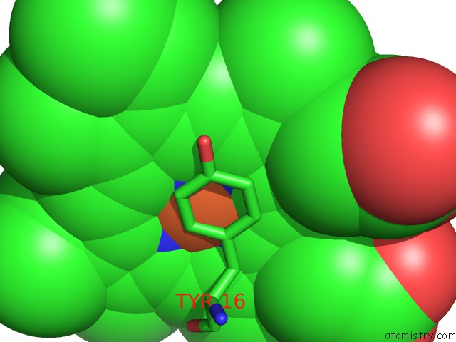
Mono view
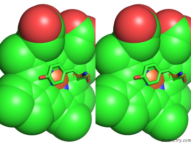
Stereo pair view
Mono view
Stereo pair view
A full contact list of Iron with other atoms in the Fe binding
site number 1 of Atomic Structure of A Cytochrome C' with An Unusual Ligand-Controlled Dimer Dissociation at 1.8 Angstroms Resolution within 5.0Å range:
|
Iron binding site 2 out of 2 in 1bbh
Go back to
Iron binding site 2 out
of 2 in the Atomic Structure of A Cytochrome C' with An Unusual Ligand-Controlled Dimer Dissociation at 1.8 Angstroms Resolution
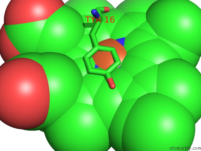
Mono view
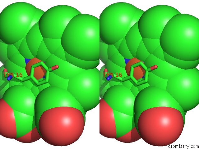
Stereo pair view
Mono view
Stereo pair view
A full contact list of Iron with other atoms in the Fe binding
site number 2 of Atomic Structure of A Cytochrome C' with An Unusual Ligand-Controlled Dimer Dissociation at 1.8 Angstroms Resolution within 5.0Å range:
|
Reference:
Z.Ren,
T.Meyer,
D.E.Mcree.
Atomic Structure of A Cytochrome C' with An Unusual Ligand-Controlled Dimer Dissociation at 1.8 A Resolution. J.Mol.Biol. V. 234 433 1993.
ISSN: ISSN 0022-2836
PubMed: 8230224
DOI: 10.1006/JMBI.1993.1597
Page generated: Wed Jul 16 12:31:18 2025
ISSN: ISSN 0022-2836
PubMed: 8230224
DOI: 10.1006/JMBI.1993.1597
Last articles
Fe in 2P17Fe in 2P0M
Fe in 2OYY
Fe in 2OZY
Fe in 2OZ0
Fe in 2OYE
Fe in 2OYU
Fe in 2OWT
Fe in 2OWJ
Fe in 2OWS