Iron »
PDB 1ch3-1cp2 »
1cno »
Iron in PDB 1cno: Structure of Pseudomonas Nautica Cytochrome C552, By Mad Method
Protein crystallography data
The structure of Structure of Pseudomonas Nautica Cytochrome C552, By Mad Method, PDB code: 1cno
was solved by
K.Brown,
D.Nurizzo,
C.Cambillau,
with X-Ray Crystallography technique. A brief refinement statistics is given in the table below:
| Resolution Low / High (Å) | 15.00 / 2.20 |
| Space group | P 31 1 2 |
| Cell size a, b, c (Å), α, β, γ (°) | 121.800, 121.800, 136.100, 90.00, 90.00, 120.00 |
| R / Rfree (%) | 17.8 / 20.8 |
Iron Binding Sites:
The binding sites of Iron atom in the Structure of Pseudomonas Nautica Cytochrome C552, By Mad Method
(pdb code 1cno). This binding sites where shown within
5.0 Angstroms radius around Iron atom.
In total 8 binding sites of Iron where determined in the Structure of Pseudomonas Nautica Cytochrome C552, By Mad Method, PDB code: 1cno:
Jump to Iron binding site number: 1; 2; 3; 4; 5; 6; 7; 8;
In total 8 binding sites of Iron where determined in the Structure of Pseudomonas Nautica Cytochrome C552, By Mad Method, PDB code: 1cno:
Jump to Iron binding site number: 1; 2; 3; 4; 5; 6; 7; 8;
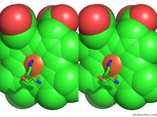
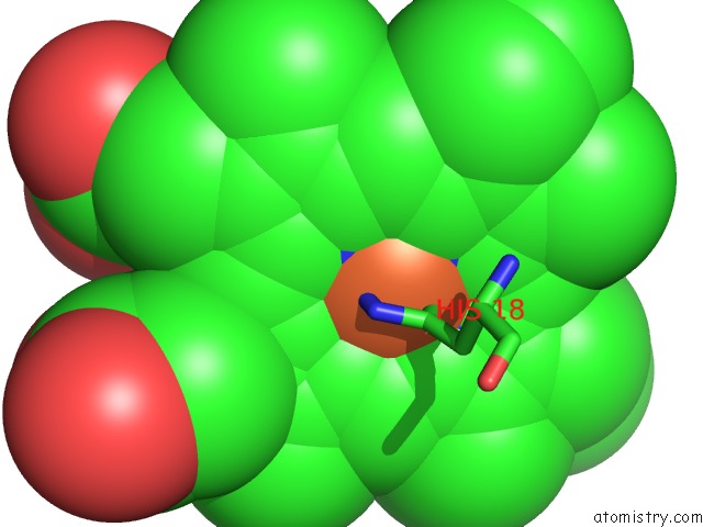
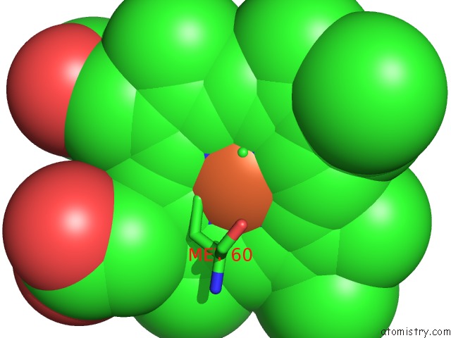
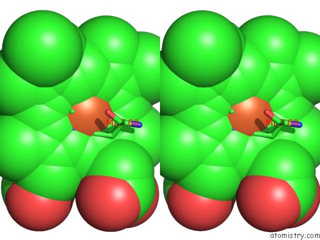
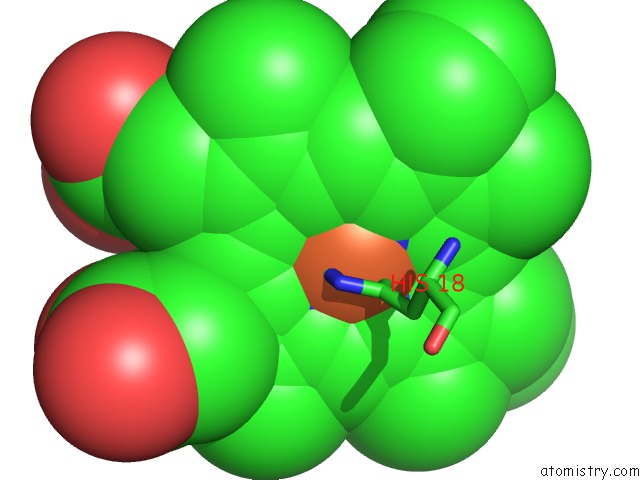
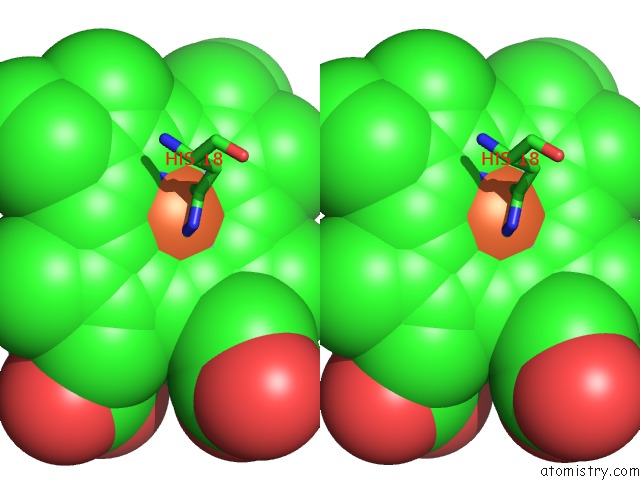
Iron binding site 1 out of 8 in 1cno
Go back to
Iron binding site 1 out
of 8 in the Structure of Pseudomonas Nautica Cytochrome C552, By Mad Method

Mono view
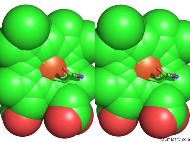
Stereo pair view
Mono view
Stereo pair view
A full contact list of Iron with other atoms in the Fe binding
site number 1 of Structure of Pseudomonas Nautica Cytochrome C552, By Mad Method within 5.0Å range:
|
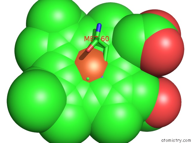
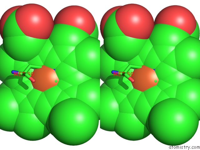
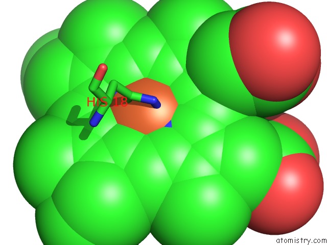
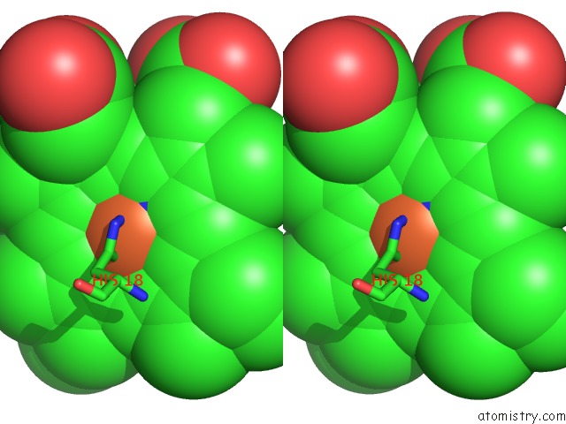
Iron binding site 2 out of 8 in 1cno
Go back to
Iron binding site 2 out
of 8 in the Structure of Pseudomonas Nautica Cytochrome C552, By Mad Method
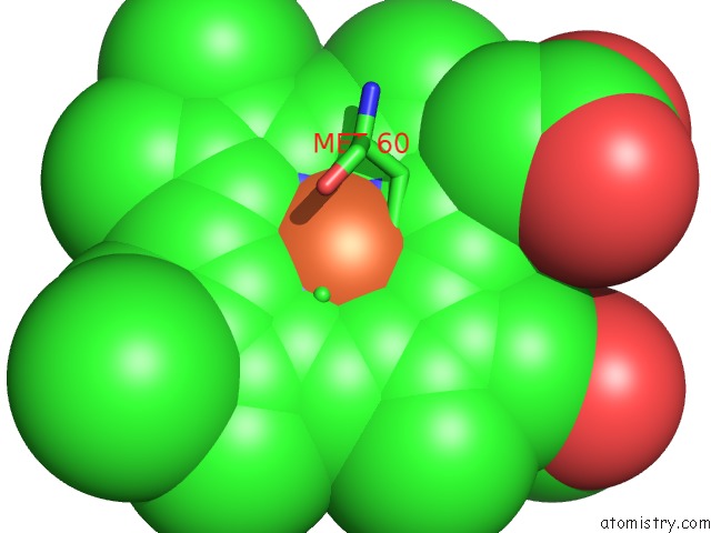
Mono view
Stereo pair view
Mono view
Stereo pair view
A full contact list of Iron with other atoms in the Fe binding
site number 2 of Structure of Pseudomonas Nautica Cytochrome C552, By Mad Method within 5.0Å range:
|
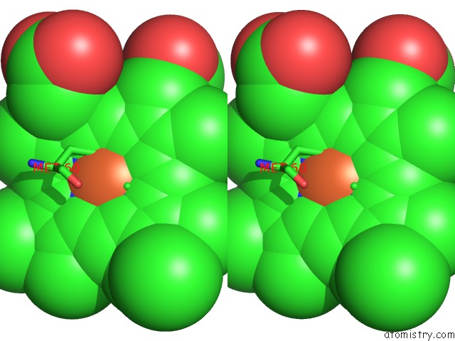
Iron binding site 3 out of 8 in 1cno
Go back to
Iron binding site 3 out
of 8 in the Structure of Pseudomonas Nautica Cytochrome C552, By Mad Method
Mono view
Stereo pair view
Mono view
Stereo pair view
A full contact list of Iron with other atoms in the Fe binding
site number 3 of Structure of Pseudomonas Nautica Cytochrome C552, By Mad Method within 5.0Å range:
|
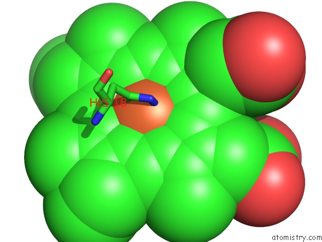
Iron binding site 4 out of 8 in 1cno
Go back to
Iron binding site 4 out
of 8 in the Structure of Pseudomonas Nautica Cytochrome C552, By Mad Method
Mono view

Stereo pair view
Mono view
Stereo pair view
A full contact list of Iron with other atoms in the Fe binding
site number 4 of Structure of Pseudomonas Nautica Cytochrome C552, By Mad Method within 5.0Å range:
|
Iron binding site 5 out of 8 in 1cno
Go back to
Iron binding site 5 out
of 8 in the Structure of Pseudomonas Nautica Cytochrome C552, By Mad Method
Mono view
Stereo pair view
Mono view
Stereo pair view
A full contact list of Iron with other atoms in the Fe binding
site number 5 of Structure of Pseudomonas Nautica Cytochrome C552, By Mad Method within 5.0Å range:
|
Iron binding site 6 out of 8 in 1cno
Go back to
Iron binding site 6 out
of 8 in the Structure of Pseudomonas Nautica Cytochrome C552, By Mad Method
Mono view
Stereo pair view
Mono view
Stereo pair view
A full contact list of Iron with other atoms in the Fe binding
site number 6 of Structure of Pseudomonas Nautica Cytochrome C552, By Mad Method within 5.0Å range:
|
Iron binding site 7 out of 8 in 1cno
Go back to
Iron binding site 7 out
of 8 in the Structure of Pseudomonas Nautica Cytochrome C552, By Mad Method
Mono view
Stereo pair view
Mono view
Stereo pair view
A full contact list of Iron with other atoms in the Fe binding
site number 7 of Structure of Pseudomonas Nautica Cytochrome C552, By Mad Method within 5.0Å range:
|
Iron binding site 8 out of 8 in 1cno
Go back to
Iron binding site 8 out
of 8 in the Structure of Pseudomonas Nautica Cytochrome C552, By Mad Method
Mono view
Stereo pair view
Mono view
Stereo pair view
A full contact list of Iron with other atoms in the Fe binding
site number 8 of Structure of Pseudomonas Nautica Cytochrome C552, By Mad Method within 5.0Å range:
|
Reference:
K.Brown,
D.Nurizzo,
S.Besson,
W.Shepard,
J.Moura,
I.Moura,
M.Tegoni,
C.Cambillau.
Mad Structure of Pseudomonas Nautica Dimeric Cytochrome C552 Mimicks the C4 Dihemic Cytochrome Domain Association. J.Mol.Biol. V. 289 1017 1999.
ISSN: ISSN 0022-2836
PubMed: 10369779
DOI: 10.1006/JMBI.1999.2838
Page generated: Wed Jul 16 13:02:21 2025
ISSN: ISSN 0022-2836
PubMed: 10369779
DOI: 10.1006/JMBI.1999.2838
Last articles
Fe in 2YXOFe in 2YRS
Fe in 2YXC
Fe in 2YNM
Fe in 2YVJ
Fe in 2YP1
Fe in 2YU2
Fe in 2YU1
Fe in 2YQB
Fe in 2YOO