Iron »
PDB 1fnq-1fz1 »
1fnq »
Iron in PDB 1fnq: Crystal Structure Analysis of the Mutant Reaction Center Pro L209-> Glu From the Photosynthetic Purple Bacterium Rhodobacter Sphaeroides
Protein crystallography data
The structure of Crystal Structure Analysis of the Mutant Reaction Center Pro L209-> Glu From the Photosynthetic Purple Bacterium Rhodobacter Sphaeroides, PDB code: 1fnq
was solved by
A.Kuglstatter,
U.Ermler,
H.Michel,
L.Baciou,
G.Fritzsch,
with X-Ray Crystallography technique. A brief refinement statistics is given in the table below:
| Resolution Low / High (Å) | 50.00 / 2.60 |
| Space group | P 31 2 1 |
| Cell size a, b, c (Å), α, β, γ (°) | 141.534, 141.534, 187.548, 90.00, 90.00, 120.00 |
| R / Rfree (%) | 21.7 / 24.7 |
Other elements in 1fnq:
The structure of Crystal Structure Analysis of the Mutant Reaction Center Pro L209-> Glu From the Photosynthetic Purple Bacterium Rhodobacter Sphaeroides also contains other interesting chemical elements:
| Magnesium | (Mg) | 4 atoms |
Iron Binding Sites:
The binding sites of Iron atom in the Crystal Structure Analysis of the Mutant Reaction Center Pro L209-> Glu From the Photosynthetic Purple Bacterium Rhodobacter Sphaeroides
(pdb code 1fnq). This binding sites where shown within
5.0 Angstroms radius around Iron atom.
In total only one binding site of Iron was determined in the Crystal Structure Analysis of the Mutant Reaction Center Pro L209-> Glu From the Photosynthetic Purple Bacterium Rhodobacter Sphaeroides, PDB code: 1fnq:
In total only one binding site of Iron was determined in the Crystal Structure Analysis of the Mutant Reaction Center Pro L209-> Glu From the Photosynthetic Purple Bacterium Rhodobacter Sphaeroides, PDB code: 1fnq:
Iron binding site 1 out of 1 in 1fnq
Go back to
Iron binding site 1 out
of 1 in the Crystal Structure Analysis of the Mutant Reaction Center Pro L209-> Glu From the Photosynthetic Purple Bacterium Rhodobacter Sphaeroides
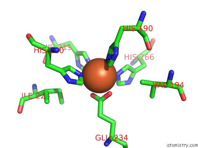
Mono view
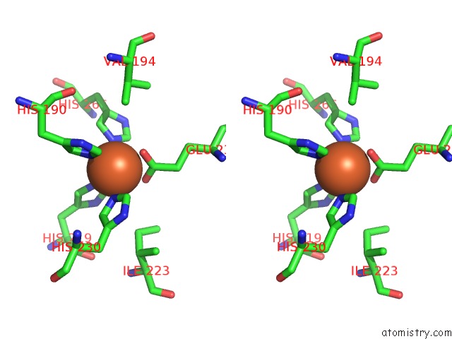
Stereo pair view
Mono view
Stereo pair view
A full contact list of Iron with other atoms in the Fe binding
site number 1 of Crystal Structure Analysis of the Mutant Reaction Center Pro L209-> Glu From the Photosynthetic Purple Bacterium Rhodobacter Sphaeroides within 5.0Å range:
|
Reference:
A.Kuglstatter,
U.Ermler,
H.Michel,
L.Baciou,
G.Fritzsch.
X-Ray Structure Analyses of Photosynthetic Reaction Center Variants From Rhodobacter Sphaeroides: Structural Changes Induced By Point Mutations at Position L209 Modulate Electron and Proton Transfer. Biochemistry V. 40 4253 2001.
ISSN: ISSN 0006-2960
PubMed: 11284681
DOI: 10.1021/BI001589H
Page generated: Sat Aug 3 05:11:20 2024
ISSN: ISSN 0006-2960
PubMed: 11284681
DOI: 10.1021/BI001589H
Last articles
Zn in 9JYWZn in 9IR4
Zn in 9IR3
Zn in 9GMX
Zn in 9GMW
Zn in 9JEJ
Zn in 9ERF
Zn in 9ERE
Zn in 9EGV
Zn in 9EGW