Iron »
PDB 1fnq-1fz1 »
1fqe »
Iron in PDB 1fqe: Crystal Structures of Mutant (K206A) That Abolish the Dilysine Interaction in the N-Lobe of Human Transferrin
Protein crystallography data
The structure of Crystal Structures of Mutant (K206A) That Abolish the Dilysine Interaction in the N-Lobe of Human Transferrin, PDB code: 1fqe
was solved by
D.Nurizzo,
H.M.Baker,
E.N.Baker,
with X-Ray Crystallography technique. A brief refinement statistics is given in the table below:
| Resolution Low / High (Å) | 14.73 / 1.80 |
| Space group | P 21 21 21 |
| Cell size a, b, c (Å), α, β, γ (°) | 43.900, 57.300, 135.900, 90.00, 90.00, 90.00 |
| R / Rfree (%) | 19.6 / 23.7 |
Other elements in 1fqe:
The structure of Crystal Structures of Mutant (K206A) That Abolish the Dilysine Interaction in the N-Lobe of Human Transferrin also contains other interesting chemical elements:
| Potassium | (K) | 1 atom |
Iron Binding Sites:
The binding sites of Iron atom in the Crystal Structures of Mutant (K206A) That Abolish the Dilysine Interaction in the N-Lobe of Human Transferrin
(pdb code 1fqe). This binding sites where shown within
5.0 Angstroms radius around Iron atom.
In total only one binding site of Iron was determined in the Crystal Structures of Mutant (K206A) That Abolish the Dilysine Interaction in the N-Lobe of Human Transferrin, PDB code: 1fqe:
In total only one binding site of Iron was determined in the Crystal Structures of Mutant (K206A) That Abolish the Dilysine Interaction in the N-Lobe of Human Transferrin, PDB code: 1fqe:
Iron binding site 1 out of 1 in 1fqe
Go back to
Iron binding site 1 out
of 1 in the Crystal Structures of Mutant (K206A) That Abolish the Dilysine Interaction in the N-Lobe of Human Transferrin
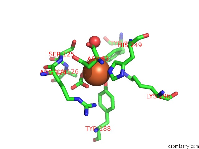
Mono view
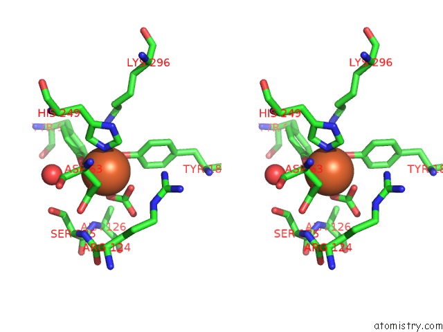
Stereo pair view
Mono view
Stereo pair view
A full contact list of Iron with other atoms in the Fe binding
site number 1 of Crystal Structures of Mutant (K206A) That Abolish the Dilysine Interaction in the N-Lobe of Human Transferrin within 5.0Å range:
|
Reference:
D.Nurizzo,
H.M.Baker,
Q.Y.He,
R.T.Macgillivray,
A.B.Mason,
R.C.Woodworth,
E.N.Baker.
Crystal Structures and Iron Release Properties of Mutants (K206A and K296A) That Abolish the Dilysine Interaction in the N-Lobe of Human Transferrin. Biochemistry V. 40 1616 2001.
ISSN: ISSN 0006-2960
PubMed: 11327820
DOI: 10.1021/BI002050M
Page generated: Wed Jul 16 14:17:28 2025
ISSN: ISSN 0006-2960
PubMed: 11327820
DOI: 10.1021/BI002050M
Last articles
Fe in 2YXOFe in 2YRS
Fe in 2YXC
Fe in 2YNM
Fe in 2YVJ
Fe in 2YP1
Fe in 2YU2
Fe in 2YU1
Fe in 2YQB
Fe in 2YOO