Iron »
PDB 1fnq-1fz1 »
1fqt »
Iron in PDB 1fqt: Crystal Structure of the Rieske-Type Ferredoxin Associated with Biphenyl Dioxygenase
Protein crystallography data
The structure of Crystal Structure of the Rieske-Type Ferredoxin Associated with Biphenyl Dioxygenase, PDB code: 1fqt
was solved by
C.L.Colbert,
M.M.-J.Couture,
L.D.Eltis,
J.T.Bolin,
with X-Ray Crystallography technique. A brief refinement statistics is given in the table below:
| Resolution Low / High (Å) | 26.00 / 1.60 |
| Space group | P 21 21 2 |
| Cell size a, b, c (Å), α, β, γ (°) | 76.400, 53.100, 64.700, 90.00, 90.00, 90.00 |
| R / Rfree (%) | 18 / 20 |
Iron Binding Sites:
The binding sites of Iron atom in the Crystal Structure of the Rieske-Type Ferredoxin Associated with Biphenyl Dioxygenase
(pdb code 1fqt). This binding sites where shown within
5.0 Angstroms radius around Iron atom.
In total 4 binding sites of Iron where determined in the Crystal Structure of the Rieske-Type Ferredoxin Associated with Biphenyl Dioxygenase, PDB code: 1fqt:
Jump to Iron binding site number: 1; 2; 3; 4;
In total 4 binding sites of Iron where determined in the Crystal Structure of the Rieske-Type Ferredoxin Associated with Biphenyl Dioxygenase, PDB code: 1fqt:
Jump to Iron binding site number: 1; 2; 3; 4;
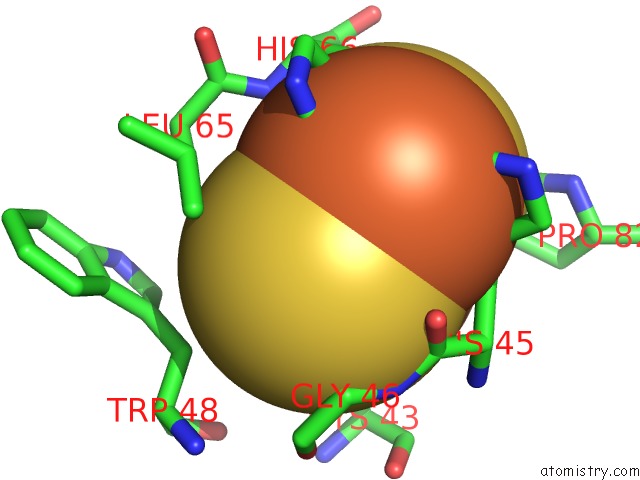
Iron binding site 1 out of 4 in 1fqt
Go back to
Iron binding site 1 out
of 4 in the Crystal Structure of the Rieske-Type Ferredoxin Associated with Biphenyl Dioxygenase
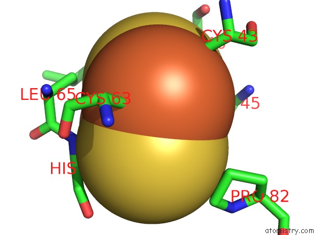
Mono view
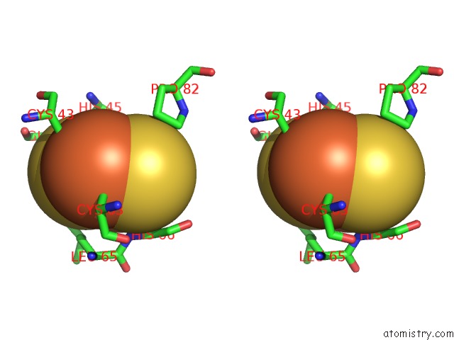
Stereo pair view
Mono view
Stereo pair view
A full contact list of Iron with other atoms in the Fe binding
site number 1 of Crystal Structure of the Rieske-Type Ferredoxin Associated with Biphenyl Dioxygenase within 5.0Å range:
|
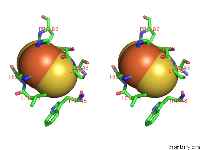
Iron binding site 2 out of 4 in 1fqt
Go back to
Iron binding site 2 out
of 4 in the Crystal Structure of the Rieske-Type Ferredoxin Associated with Biphenyl Dioxygenase
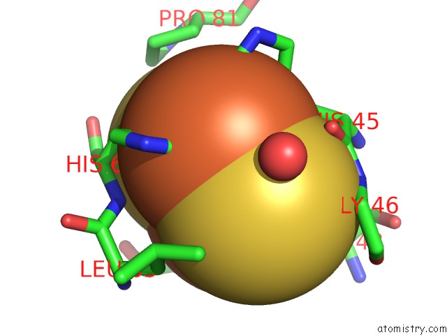
Mono view
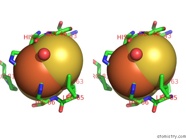
Stereo pair view
Mono view
Stereo pair view
A full contact list of Iron with other atoms in the Fe binding
site number 2 of Crystal Structure of the Rieske-Type Ferredoxin Associated with Biphenyl Dioxygenase within 5.0Å range:
|
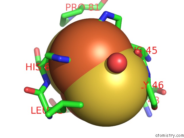
Iron binding site 3 out of 4 in 1fqt
Go back to
Iron binding site 3 out
of 4 in the Crystal Structure of the Rieske-Type Ferredoxin Associated with Biphenyl Dioxygenase
Mono view
Stereo pair view
Mono view
Stereo pair view
A full contact list of Iron with other atoms in the Fe binding
site number 3 of Crystal Structure of the Rieske-Type Ferredoxin Associated with Biphenyl Dioxygenase within 5.0Å range:
|
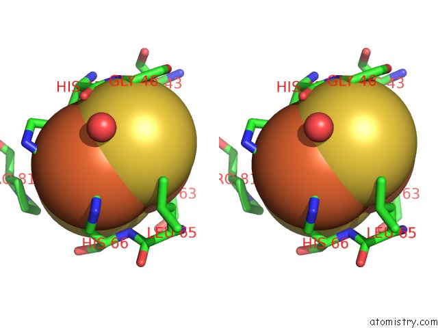
Iron binding site 4 out of 4 in 1fqt
Go back to
Iron binding site 4 out
of 4 in the Crystal Structure of the Rieske-Type Ferredoxin Associated with Biphenyl Dioxygenase
Mono view
Stereo pair view
Mono view
Stereo pair view
A full contact list of Iron with other atoms in the Fe binding
site number 4 of Crystal Structure of the Rieske-Type Ferredoxin Associated with Biphenyl Dioxygenase within 5.0Å range:
|
Reference:
C.L.Colbert,
M.M.Couture,
L.D.Eltis,
J.T.Bolin.
A Cluster Exposed: Structure of the Rieske Ferredoxin From Biphenyl Dioxygenase and the Redox Properties of Rieske Fe-S Proteins. Structure Fold.Des. V. 8 1267 2000.
ISSN: ISSN 0969-2126
PubMed: 11188691
DOI: 10.1016/S0969-2126(00)00536-0
Page generated: Sat Aug 3 05:13:23 2024
ISSN: ISSN 0969-2126
PubMed: 11188691
DOI: 10.1016/S0969-2126(00)00536-0
Last articles
F in 7Q2JF in 7Q01
F in 7PZX
F in 7PZW
F in 7PZV
F in 7PZU
F in 7PZS
F in 7PY4
F in 7PX6
F in 7PVU