Iron »
PDB 1fnq-1fz1 »
1fxd »
Iron in PDB 1fxd: Refined Crystal Structure of Ferredoxin II From Desulfovibrio Gigas at 1.7 Angstroms
Protein crystallography data
The structure of Refined Crystal Structure of Ferredoxin II From Desulfovibrio Gigas at 1.7 Angstroms, PDB code: 1fxd
was solved by
C.R.Kissinger,
L.C.Sieker,
E.T.Adman,
L.H.Jensen,
with X-Ray Crystallography technique. A brief refinement statistics is given in the table below:
| Resolution Low / High (Å) | N/A / 1.70 |
| Space group | C 1 2 1 |
| Cell size a, b, c (Å), α, β, γ (°) | 40.870, 45.280, 26.470, 90.00, 104.70, 90.00 |
| R / Rfree (%) | n/a / n/a |
Iron Binding Sites:
The binding sites of Iron atom in the Refined Crystal Structure of Ferredoxin II From Desulfovibrio Gigas at 1.7 Angstroms
(pdb code 1fxd). This binding sites where shown within
5.0 Angstroms radius around Iron atom.
In total 3 binding sites of Iron where determined in the Refined Crystal Structure of Ferredoxin II From Desulfovibrio Gigas at 1.7 Angstroms, PDB code: 1fxd:
Jump to Iron binding site number: 1; 2; 3;
In total 3 binding sites of Iron where determined in the Refined Crystal Structure of Ferredoxin II From Desulfovibrio Gigas at 1.7 Angstroms, PDB code: 1fxd:
Jump to Iron binding site number: 1; 2; 3;
Iron binding site 1 out of 3 in 1fxd
Go back to
Iron binding site 1 out
of 3 in the Refined Crystal Structure of Ferredoxin II From Desulfovibrio Gigas at 1.7 Angstroms

Mono view
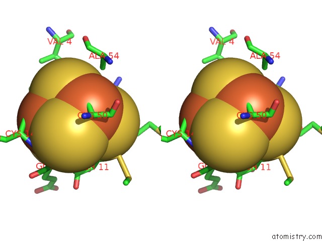
Stereo pair view
Mono view
Stereo pair view
A full contact list of Iron with other atoms in the Fe binding
site number 1 of Refined Crystal Structure of Ferredoxin II From Desulfovibrio Gigas at 1.7 Angstroms within 5.0Å range:
|
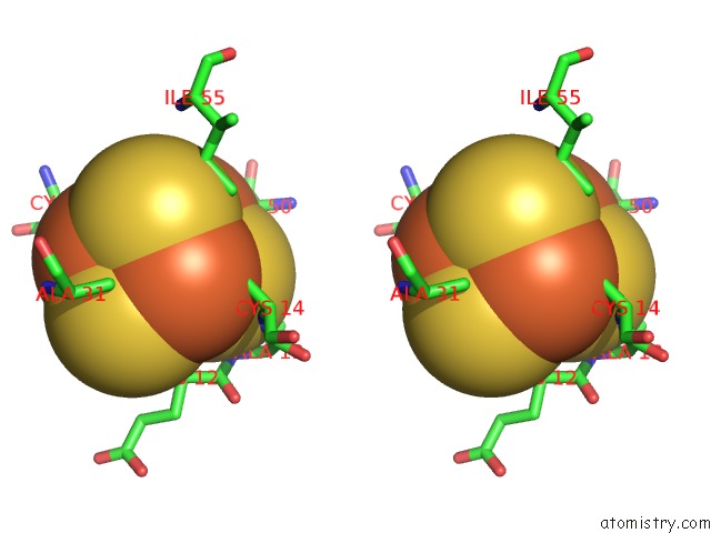
Iron binding site 2 out of 3 in 1fxd
Go back to
Iron binding site 2 out
of 3 in the Refined Crystal Structure of Ferredoxin II From Desulfovibrio Gigas at 1.7 Angstroms

Mono view
Stereo pair view
Mono view
Stereo pair view
A full contact list of Iron with other atoms in the Fe binding
site number 2 of Refined Crystal Structure of Ferredoxin II From Desulfovibrio Gigas at 1.7 Angstroms within 5.0Å range:
|
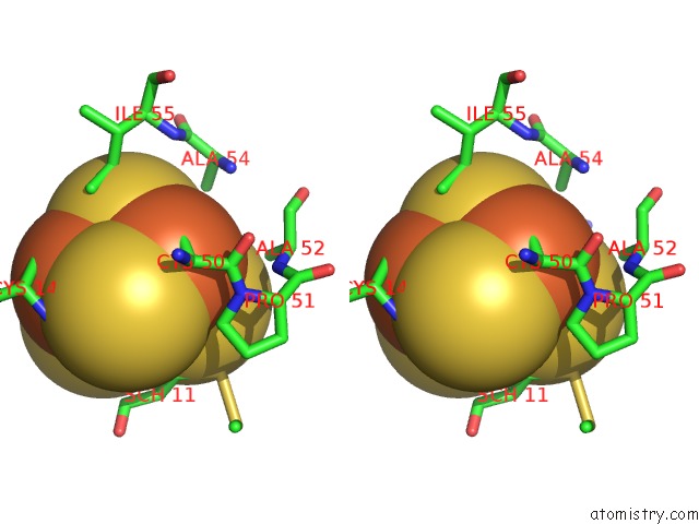
Iron binding site 3 out of 3 in 1fxd
Go back to
Iron binding site 3 out
of 3 in the Refined Crystal Structure of Ferredoxin II From Desulfovibrio Gigas at 1.7 Angstroms
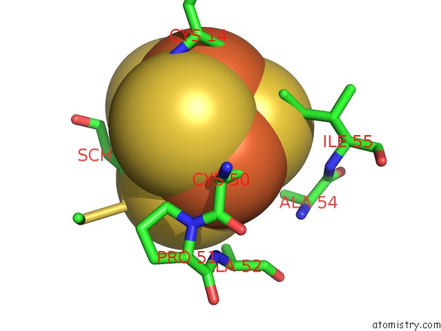
Mono view
Stereo pair view
Mono view
Stereo pair view
A full contact list of Iron with other atoms in the Fe binding
site number 3 of Refined Crystal Structure of Ferredoxin II From Desulfovibrio Gigas at 1.7 Angstroms within 5.0Å range:
|
Reference:
C.R.Kissinger,
L.C.Sieker,
E.T.Adman,
L.H.Jensen.
Refined Crystal Structure of Ferredoxin II From Desulfovibrio Gigas at 1.7 A. J.Mol.Biol. V. 219 693 1991.
ISSN: ISSN 0022-2836
PubMed: 2056535
DOI: 10.1016/0022-2836(91)90665-S
Page generated: Sat Aug 3 05:29:37 2024
ISSN: ISSN 0022-2836
PubMed: 2056535
DOI: 10.1016/0022-2836(91)90665-S
Last articles
Zn in 9J0NZn in 9J0O
Zn in 9J0P
Zn in 9FJX
Zn in 9EKB
Zn in 9C0F
Zn in 9CAH
Zn in 9CH0
Zn in 9CH3
Zn in 9CH1