Iron »
PDB 1gem-1gwe »
1gge »
Iron in PDB 1gge: Crystal Structure of Catalase Hpii From Escherichia Coli, Native Structure at 1.9 A Resolution.
Enzymatic activity of Crystal Structure of Catalase Hpii From Escherichia Coli, Native Structure at 1.9 A Resolution.
All present enzymatic activity of Crystal Structure of Catalase Hpii From Escherichia Coli, Native Structure at 1.9 A Resolution.:
1.11.1.6;
1.11.1.6;
Protein crystallography data
The structure of Crystal Structure of Catalase Hpii From Escherichia Coli, Native Structure at 1.9 A Resolution., PDB code: 1gge
was solved by
W.R.Melik-Adamyan,
J.Bravo,
X.Carpena,
J.Switala,
M.J.Mate,
I.Fita,
P.C.Loewen,
with X-Ray Crystallography technique. A brief refinement statistics is given in the table below:
| Resolution Low / High (Å) | 87.60 / 1.89 |
| Space group | P 1 21 1 |
| Cell size a, b, c (Å), α, β, γ (°) | 93.040, 132.340, 121.200, 90.00, 109.63, 90.00 |
| R / Rfree (%) | 16.3 / 20.2 |
Iron Binding Sites:
The binding sites of Iron atom in the Crystal Structure of Catalase Hpii From Escherichia Coli, Native Structure at 1.9 A Resolution.
(pdb code 1gge). This binding sites where shown within
5.0 Angstroms radius around Iron atom.
In total 4 binding sites of Iron where determined in the Crystal Structure of Catalase Hpii From Escherichia Coli, Native Structure at 1.9 A Resolution., PDB code: 1gge:
Jump to Iron binding site number: 1; 2; 3; 4;
In total 4 binding sites of Iron where determined in the Crystal Structure of Catalase Hpii From Escherichia Coli, Native Structure at 1.9 A Resolution., PDB code: 1gge:
Jump to Iron binding site number: 1; 2; 3; 4;
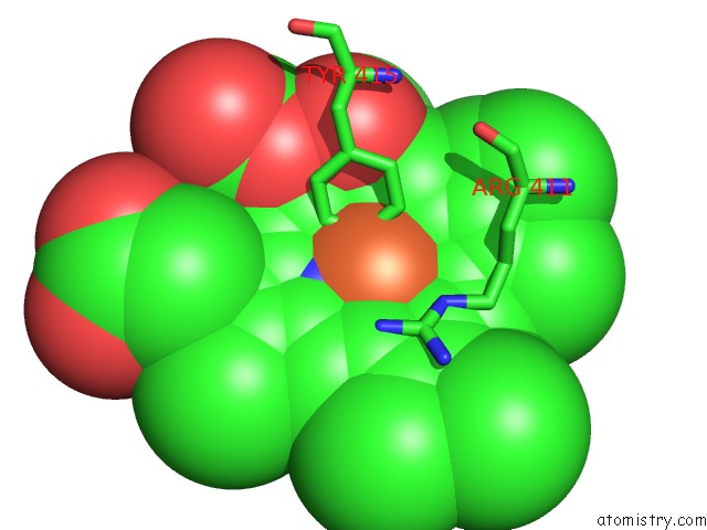
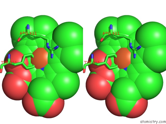
Iron binding site 1 out of 4 in 1gge
Go back to
Iron binding site 1 out
of 4 in the Crystal Structure of Catalase Hpii From Escherichia Coli, Native Structure at 1.9 A Resolution.
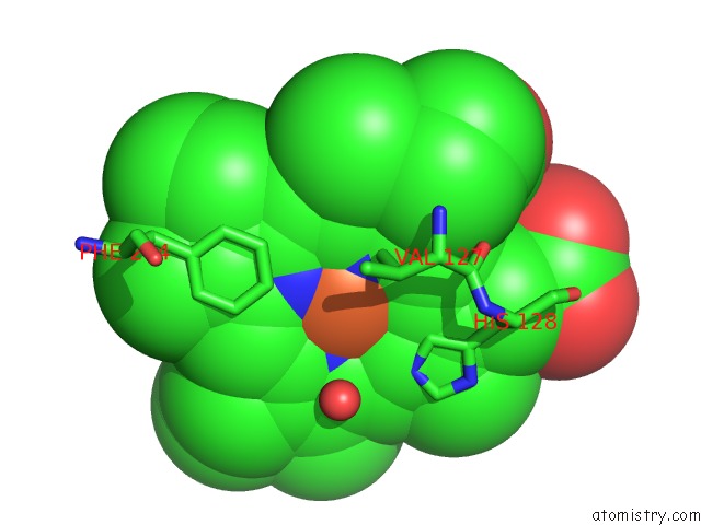
Mono view
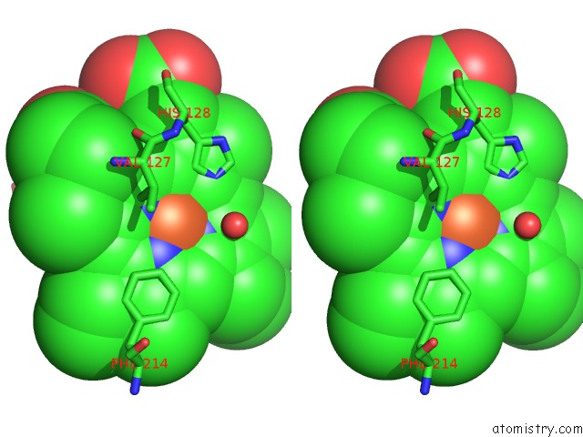
Stereo pair view
Mono view
Stereo pair view
A full contact list of Iron with other atoms in the Fe binding
site number 1 of Crystal Structure of Catalase Hpii From Escherichia Coli, Native Structure at 1.9 A Resolution. within 5.0Å range:
|
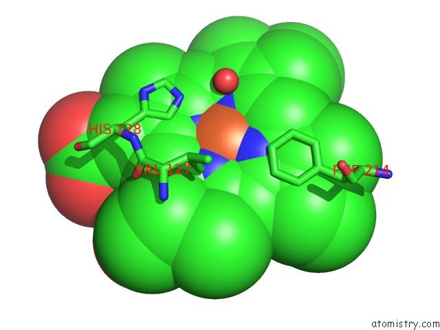
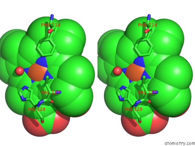
Iron binding site 2 out of 4 in 1gge
Go back to
Iron binding site 2 out
of 4 in the Crystal Structure of Catalase Hpii From Escherichia Coli, Native Structure at 1.9 A Resolution.
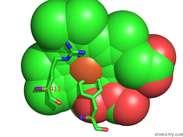
Mono view
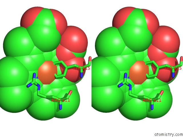
Stereo pair view
Mono view
Stereo pair view
A full contact list of Iron with other atoms in the Fe binding
site number 2 of Crystal Structure of Catalase Hpii From Escherichia Coli, Native Structure at 1.9 A Resolution. within 5.0Å range:
|
Iron binding site 3 out of 4 in 1gge
Go back to
Iron binding site 3 out
of 4 in the Crystal Structure of Catalase Hpii From Escherichia Coli, Native Structure at 1.9 A Resolution.
Mono view
Stereo pair view
Mono view
Stereo pair view
A full contact list of Iron with other atoms in the Fe binding
site number 3 of Crystal Structure of Catalase Hpii From Escherichia Coli, Native Structure at 1.9 A Resolution. within 5.0Å range:
|
Iron binding site 4 out of 4 in 1gge
Go back to
Iron binding site 4 out
of 4 in the Crystal Structure of Catalase Hpii From Escherichia Coli, Native Structure at 1.9 A Resolution.
Mono view
Stereo pair view
Mono view
Stereo pair view
A full contact list of Iron with other atoms in the Fe binding
site number 4 of Crystal Structure of Catalase Hpii From Escherichia Coli, Native Structure at 1.9 A Resolution. within 5.0Å range:
|
Reference:
W.Melik-Adamyan,
J.Bravo,
X.Carpena,
J.Switala,
M.J.Mate,
I.Fita,
P.C.Loewen.
Substrate Flow in Catalases Deduced From the Crystal Structures of Active Site Variants of Hpii From Escherichia Coli. Proteins V. 44 270 2001.
ISSN: ISSN 0887-3585
PubMed: 11455600
DOI: 10.1002/PROT.1092
Page generated: Sat Aug 3 06:09:48 2024
ISSN: ISSN 0887-3585
PubMed: 11455600
DOI: 10.1002/PROT.1092
Last articles
Zn in 9J0NZn in 9J0O
Zn in 9J0P
Zn in 9FJX
Zn in 9EKB
Zn in 9C0F
Zn in 9CAH
Zn in 9CH0
Zn in 9CH3
Zn in 9CH1