Iron »
PDB 1gem-1gwe »
1gli »
Iron in PDB 1gli: Deoxyhemoglobin T38W (Alpha Chains), V1G (Alpha and Beta Chains)
Protein crystallography data
The structure of Deoxyhemoglobin T38W (Alpha Chains), V1G (Alpha and Beta Chains), PDB code: 1gli
was solved by
G.Fermi,
B.Vallone,
with X-Ray Crystallography technique. A brief refinement statistics is given in the table below:
| Resolution Low / High (Å) | N/A / 2.50 |
| Space group | P 1 21 1 |
| Cell size a, b, c (Å), α, β, γ (°) | 63.150, 83.590, 53.800, 90.00, 99.34, 90.00 |
| R / Rfree (%) | n/a / n/a |
Iron Binding Sites:
The binding sites of Iron atom in the Deoxyhemoglobin T38W (Alpha Chains), V1G (Alpha and Beta Chains)
(pdb code 1gli). This binding sites where shown within
5.0 Angstroms radius around Iron atom.
In total 4 binding sites of Iron where determined in the Deoxyhemoglobin T38W (Alpha Chains), V1G (Alpha and Beta Chains), PDB code: 1gli:
Jump to Iron binding site number: 1; 2; 3; 4;
In total 4 binding sites of Iron where determined in the Deoxyhemoglobin T38W (Alpha Chains), V1G (Alpha and Beta Chains), PDB code: 1gli:
Jump to Iron binding site number: 1; 2; 3; 4;
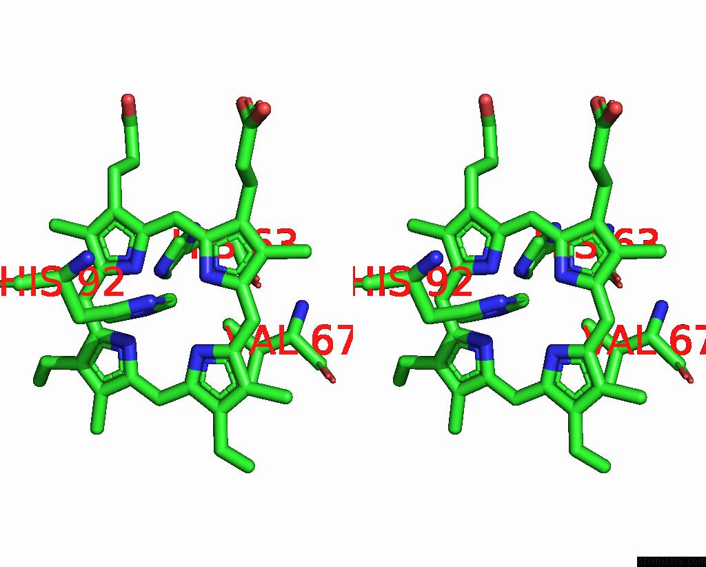
Iron binding site 1 out of 4 in 1gli
Go back to
Iron binding site 1 out
of 4 in the Deoxyhemoglobin T38W (Alpha Chains), V1G (Alpha and Beta Chains)

Mono view
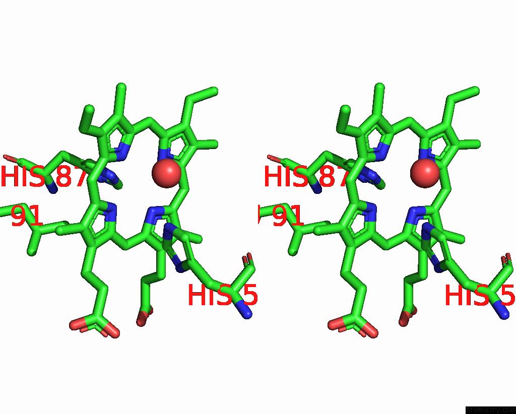
Stereo pair view
Mono view
Stereo pair view
A full contact list of Iron with other atoms in the Fe binding
site number 1 of Deoxyhemoglobin T38W (Alpha Chains), V1G (Alpha and Beta Chains) within 5.0Å range:
|
Iron binding site 2 out of 4 in 1gli
Go back to
Iron binding site 2 out
of 4 in the Deoxyhemoglobin T38W (Alpha Chains), V1G (Alpha and Beta Chains)

Mono view
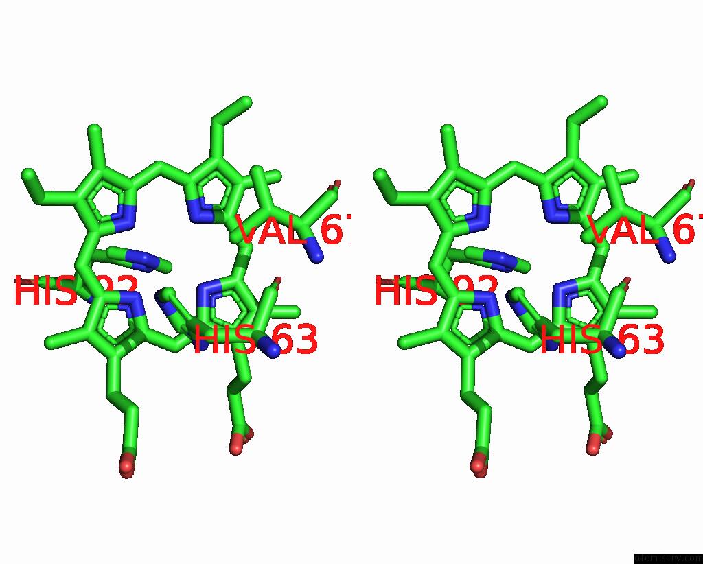
Stereo pair view
Mono view
Stereo pair view
A full contact list of Iron with other atoms in the Fe binding
site number 2 of Deoxyhemoglobin T38W (Alpha Chains), V1G (Alpha and Beta Chains) within 5.0Å range:
|
Iron binding site 3 out of 4 in 1gli
Go back to
Iron binding site 3 out
of 4 in the Deoxyhemoglobin T38W (Alpha Chains), V1G (Alpha and Beta Chains)

Mono view
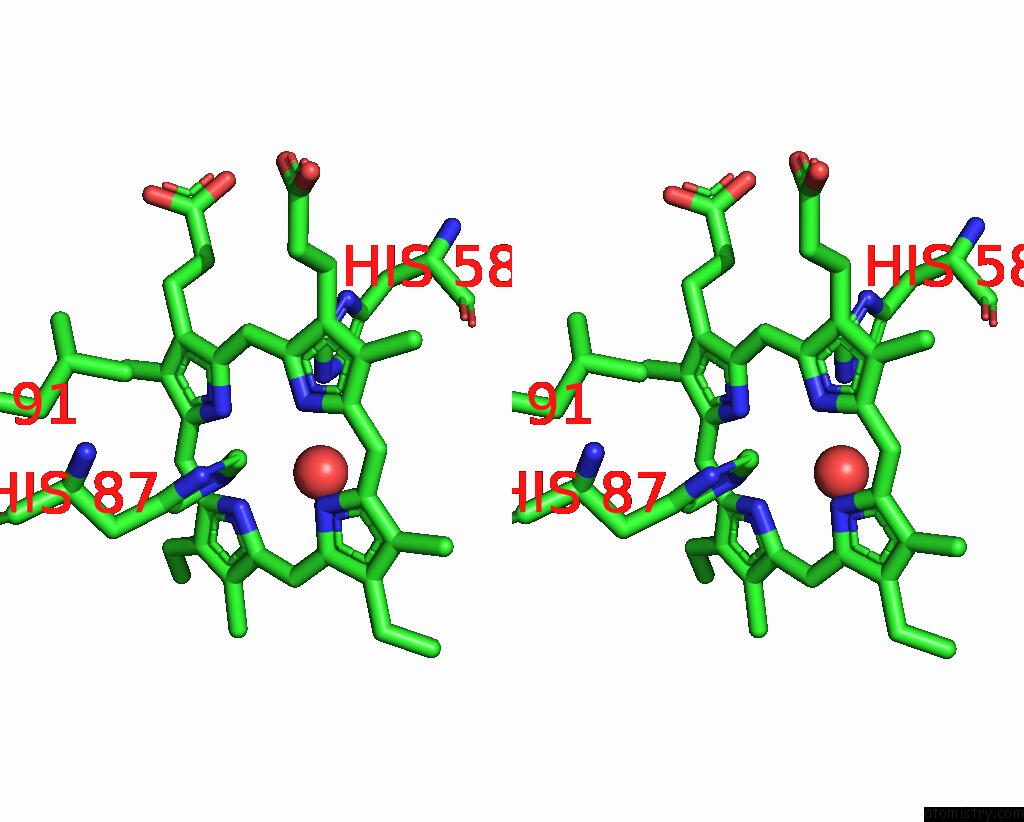
Stereo pair view
Mono view
Stereo pair view
A full contact list of Iron with other atoms in the Fe binding
site number 3 of Deoxyhemoglobin T38W (Alpha Chains), V1G (Alpha and Beta Chains) within 5.0Å range:
|
Iron binding site 4 out of 4 in 1gli
Go back to
Iron binding site 4 out
of 4 in the Deoxyhemoglobin T38W (Alpha Chains), V1G (Alpha and Beta Chains)
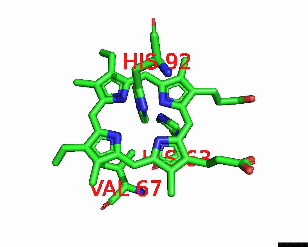
Mono view
Stereo pair view
Mono view
Stereo pair view
A full contact list of Iron with other atoms in the Fe binding
site number 4 of Deoxyhemoglobin T38W (Alpha Chains), V1G (Alpha and Beta Chains) within 5.0Å range:
|
Reference:
B.Vallone,
A.Bellelli,
A.E.Miele,
M.Brunori,
G.Fermi.
Probing the Alpha 1 Beta 2 Interface of Human Hemoglobin By Mutagenesis. Role of the Fg-C Contact Regions. J.Biol.Chem. V. 271 12472 1996.
ISSN: ISSN 0021-9258
PubMed: 8647854
DOI: 10.1074/JBC.271.21.12472
Page generated: Sat Aug 3 06:11:03 2024
ISSN: ISSN 0021-9258
PubMed: 8647854
DOI: 10.1074/JBC.271.21.12472
Last articles
Zn in 9J0NZn in 9J0O
Zn in 9J0P
Zn in 9FJX
Zn in 9EKB
Zn in 9C0F
Zn in 9CAH
Zn in 9CH0
Zn in 9CH3
Zn in 9CH1