Iron »
PDB 1gem-1gwe »
1guq »
Iron in PDB 1guq: Structure of Nucleotidyltransferase Complexed with Udp-Glucose
Enzymatic activity of Structure of Nucleotidyltransferase Complexed with Udp-Glucose
All present enzymatic activity of Structure of Nucleotidyltransferase Complexed with Udp-Glucose:
2.7.7.10;
2.7.7.10;
Protein crystallography data
The structure of Structure of Nucleotidyltransferase Complexed with Udp-Glucose, PDB code: 1guq
was solved by
J.B.Thoden,
I.Rayment,
H.Holden,
with X-Ray Crystallography technique. A brief refinement statistics is given in the table below:
| Resolution Low / High (Å) | 30.00 / 1.80 |
| Space group | P 1 21 1 |
| Cell size a, b, c (Å), α, β, γ (°) | 68.400, 57.500, 188.900, 90.00, 100.13, 90.00 |
| R / Rfree (%) | n/a / n/a |
Other elements in 1guq:
The structure of Structure of Nucleotidyltransferase Complexed with Udp-Glucose also contains other interesting chemical elements:
| Potassium | (K) | 4 atoms |
| Zinc | (Zn) | 4 atoms |
Iron Binding Sites:
The binding sites of Iron atom in the Structure of Nucleotidyltransferase Complexed with Udp-Glucose
(pdb code 1guq). This binding sites where shown within
5.0 Angstroms radius around Iron atom.
In total 4 binding sites of Iron where determined in the Structure of Nucleotidyltransferase Complexed with Udp-Glucose, PDB code: 1guq:
Jump to Iron binding site number: 1; 2; 3; 4;
In total 4 binding sites of Iron where determined in the Structure of Nucleotidyltransferase Complexed with Udp-Glucose, PDB code: 1guq:
Jump to Iron binding site number: 1; 2; 3; 4;
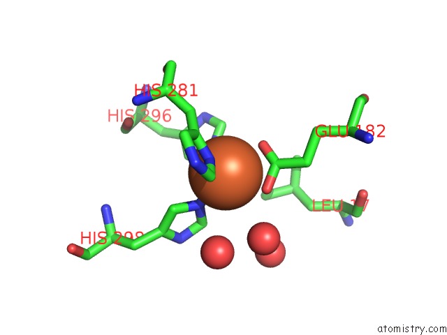
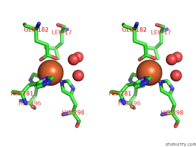
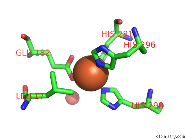
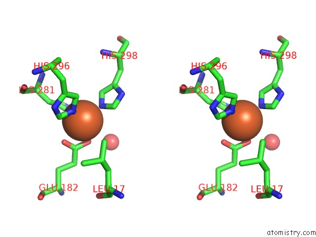
Iron binding site 1 out of 4 in 1guq
Go back to
Iron binding site 1 out
of 4 in the Structure of Nucleotidyltransferase Complexed with Udp-Glucose
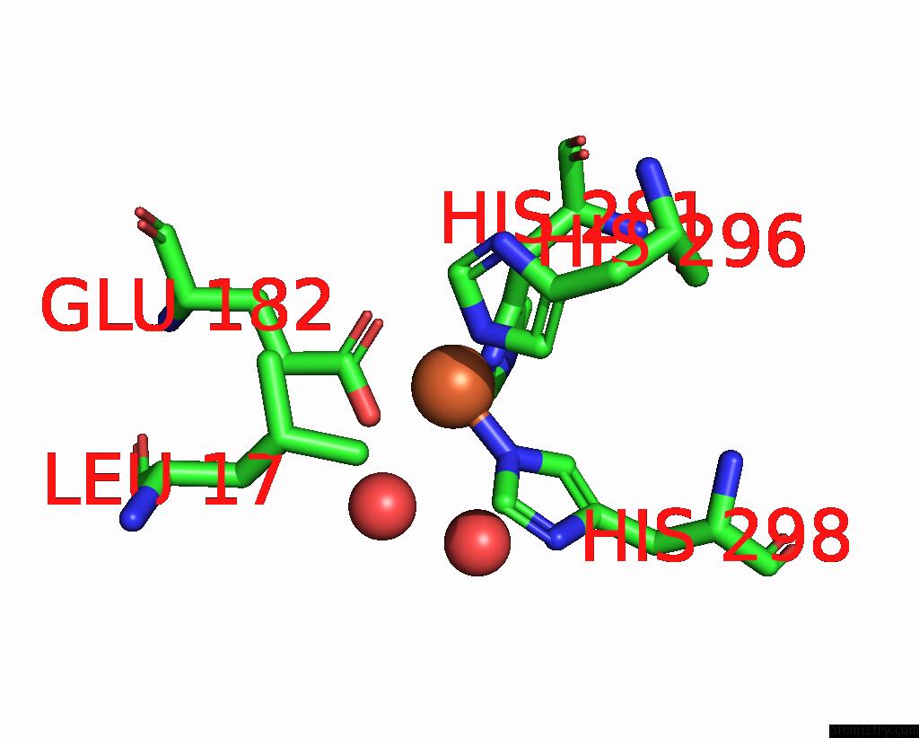
Mono view
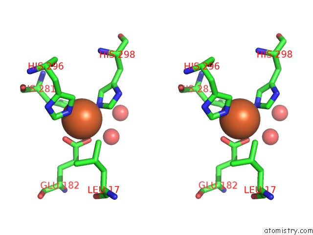
Stereo pair view
Mono view
Stereo pair view
A full contact list of Iron with other atoms in the Fe binding
site number 1 of Structure of Nucleotidyltransferase Complexed with Udp-Glucose within 5.0Å range:
|
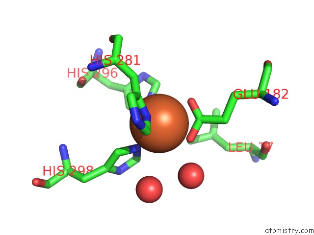
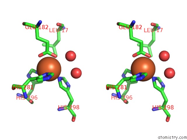
Iron binding site 2 out of 4 in 1guq
Go back to
Iron binding site 2 out
of 4 in the Structure of Nucleotidyltransferase Complexed with Udp-Glucose
Mono view
Stereo pair view
Mono view
Stereo pair view
A full contact list of Iron with other atoms in the Fe binding
site number 2 of Structure of Nucleotidyltransferase Complexed with Udp-Glucose within 5.0Å range:
|
Iron binding site 3 out of 4 in 1guq
Go back to
Iron binding site 3 out
of 4 in the Structure of Nucleotidyltransferase Complexed with Udp-Glucose
Mono view
Stereo pair view
Mono view
Stereo pair view
A full contact list of Iron with other atoms in the Fe binding
site number 3 of Structure of Nucleotidyltransferase Complexed with Udp-Glucose within 5.0Å range:
|
Iron binding site 4 out of 4 in 1guq
Go back to
Iron binding site 4 out
of 4 in the Structure of Nucleotidyltransferase Complexed with Udp-Glucose
Mono view
Stereo pair view
Mono view
Stereo pair view
A full contact list of Iron with other atoms in the Fe binding
site number 4 of Structure of Nucleotidyltransferase Complexed with Udp-Glucose within 5.0Å range:
|
Reference:
J.B.Thoden,
F.J.Ruzicka,
P.A.Frey,
I.Rayment,
H.M.Holden.
Structural Analysis of the H166G Site-Directed Mutant of Galactose-1-Phosphate Uridylyltransferase Complexed with Either Udp-Glucose or Udp-Galactose: Detailed Description of the Nucleotide Sugar Binding Site. Biochemistry V. 36 1212 1997.
ISSN: ISSN 0006-2960
PubMed: 9063869
DOI: 10.1021/BI9626517
Page generated: Wed Jul 16 15:15:47 2025
ISSN: ISSN 0006-2960
PubMed: 9063869
DOI: 10.1021/BI9626517
Last articles
Fe in 2YXOFe in 2YRS
Fe in 2YXC
Fe in 2YNM
Fe in 2YVJ
Fe in 2YP1
Fe in 2YU2
Fe in 2YU1
Fe in 2YQB
Fe in 2YOO