Iron »
PDB 1hzu-1ird »
1ibe »
Iron in PDB 1ibe: Deoxy-Haemoglobin Trapped in the High-Affinity (R) State
Protein crystallography data
The structure of Deoxy-Haemoglobin Trapped in the High-Affinity (R) State, PDB code: 1ibe
was solved by
J.Wilson,
K.Phillips,
B.Luisi,
with X-Ray Crystallography technique. A brief refinement statistics is given in the table below:
| Resolution Low / High (Å) | N/A / 1.80 |
| Space group | C 1 2 1 |
| Cell size a, b, c (Å), α, β, γ (°) | 108.180, 63.180, 54.580, 90.00, 111.10, 90.00 |
| R / Rfree (%) | n/a / n/a |
Iron Binding Sites:
The binding sites of Iron atom in the Deoxy-Haemoglobin Trapped in the High-Affinity (R) State
(pdb code 1ibe). This binding sites where shown within
5.0 Angstroms radius around Iron atom.
In total 2 binding sites of Iron where determined in the Deoxy-Haemoglobin Trapped in the High-Affinity (R) State, PDB code: 1ibe:
Jump to Iron binding site number: 1; 2;
In total 2 binding sites of Iron where determined in the Deoxy-Haemoglobin Trapped in the High-Affinity (R) State, PDB code: 1ibe:
Jump to Iron binding site number: 1; 2;
Iron binding site 1 out of 2 in 1ibe
Go back to
Iron binding site 1 out
of 2 in the Deoxy-Haemoglobin Trapped in the High-Affinity (R) State
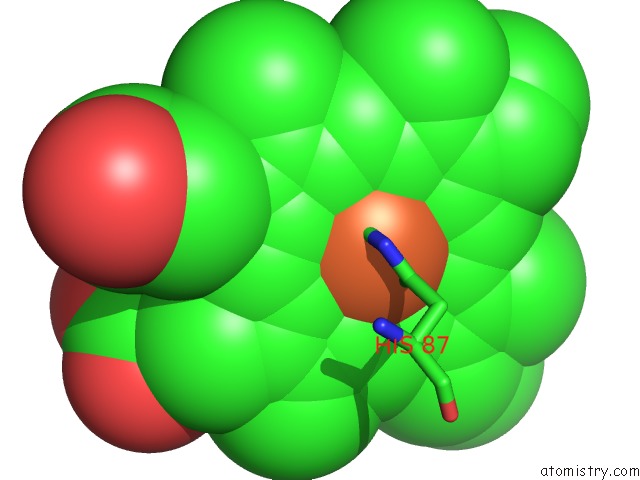
Mono view
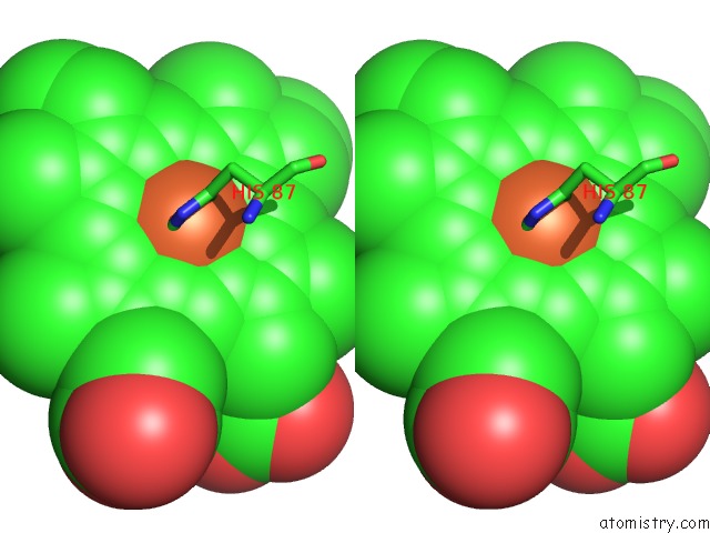
Stereo pair view
Mono view
Stereo pair view
A full contact list of Iron with other atoms in the Fe binding
site number 1 of Deoxy-Haemoglobin Trapped in the High-Affinity (R) State within 5.0Å range:
|
Iron binding site 2 out of 2 in 1ibe
Go back to
Iron binding site 2 out
of 2 in the Deoxy-Haemoglobin Trapped in the High-Affinity (R) State
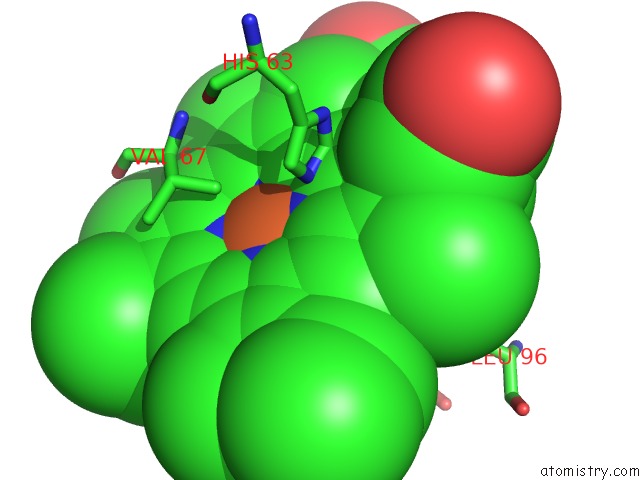
Mono view
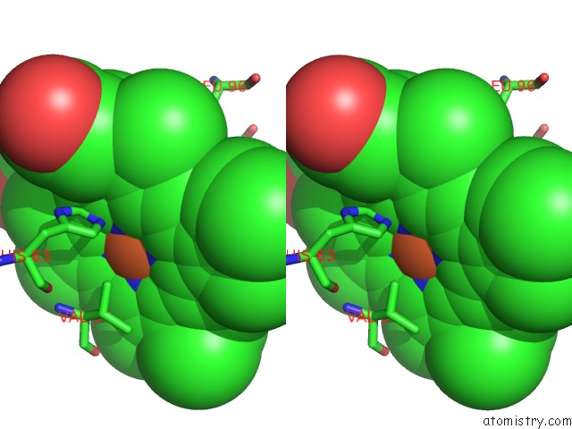
Stereo pair view
Mono view
Stereo pair view
A full contact list of Iron with other atoms in the Fe binding
site number 2 of Deoxy-Haemoglobin Trapped in the High-Affinity (R) State within 5.0Å range:
|
Reference:
J.Wilson,
K.Phillips,
B.Luisi.
The Crystal Structure of Horse Deoxyhaemoglobin Trapped in the High-Affinity (R) State. J.Mol.Biol. V. 264 743 1996.
ISSN: ISSN 0022-2836
PubMed: 8980683
DOI: 10.1006/JMBI.1996.0674
Page generated: Sat Aug 3 08:02:24 2024
ISSN: ISSN 0022-2836
PubMed: 8980683
DOI: 10.1006/JMBI.1996.0674
Last articles
Zn in 9J0NZn in 9J0O
Zn in 9J0P
Zn in 9FJX
Zn in 9EKB
Zn in 9C0F
Zn in 9CAH
Zn in 9CH0
Zn in 9CH3
Zn in 9CH1