Iron »
PDB 1j3z-1jju »
1j7y »
Iron in PDB 1j7y: Crystal Structure of Partially Ligated Mutant of Hba
Protein crystallography data
The structure of Crystal Structure of Partially Ligated Mutant of Hba, PDB code: 1j7y
was solved by
A.E.Miele,
F.Draghi,
A.Arcovito,
A.Bellelli,
M.Brunori,
C.Travaglini-Allocatelli,
B.Vallone,
with X-Ray Crystallography technique. A brief refinement statistics is given in the table below:
| Resolution Low / High (Å) | 14.90 / 1.70 |
| Space group | P 1 21 1 |
| Cell size a, b, c (Å), α, β, γ (°) | 62.570, 82.470, 53.690, 90.00, 99.70, 90.00 |
| R / Rfree (%) | 15.5 / 20.1 |
Iron Binding Sites:
The binding sites of Iron atom in the Crystal Structure of Partially Ligated Mutant of Hba
(pdb code 1j7y). This binding sites where shown within
5.0 Angstroms radius around Iron atom.
In total 4 binding sites of Iron where determined in the Crystal Structure of Partially Ligated Mutant of Hba, PDB code: 1j7y:
Jump to Iron binding site number: 1; 2; 3; 4;
In total 4 binding sites of Iron where determined in the Crystal Structure of Partially Ligated Mutant of Hba, PDB code: 1j7y:
Jump to Iron binding site number: 1; 2; 3; 4;
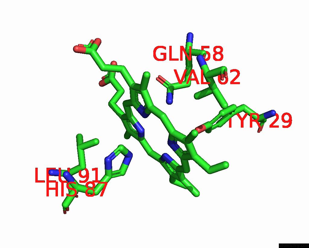
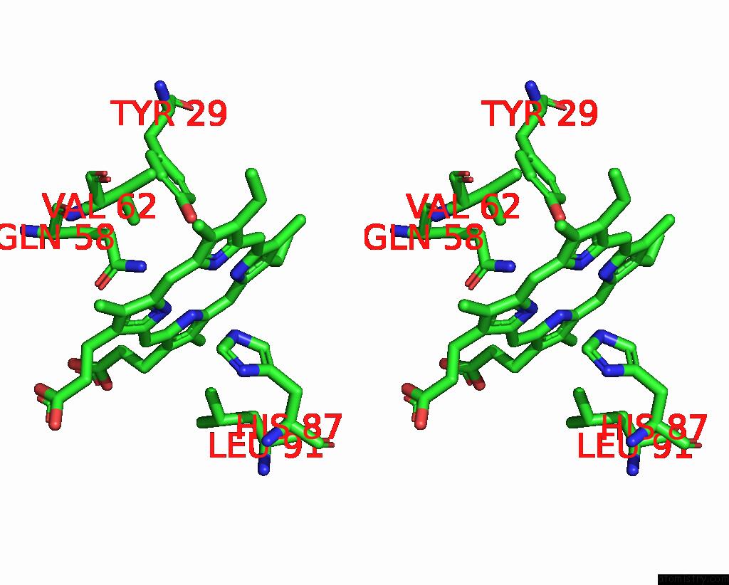
Iron binding site 1 out of 4 in 1j7y
Go back to
Iron binding site 1 out
of 4 in the Crystal Structure of Partially Ligated Mutant of Hba
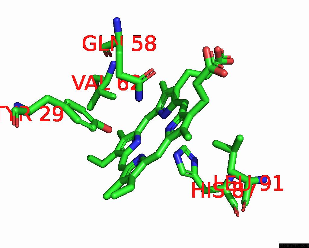
Mono view
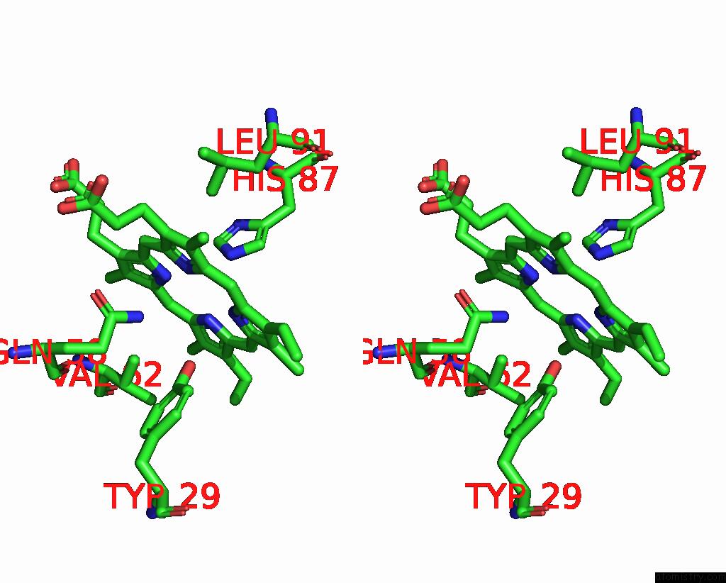
Stereo pair view
Mono view
Stereo pair view
A full contact list of Iron with other atoms in the Fe binding
site number 1 of Crystal Structure of Partially Ligated Mutant of Hba within 5.0Å range:
|
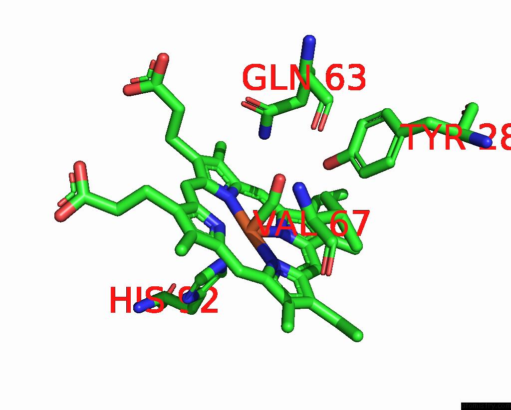
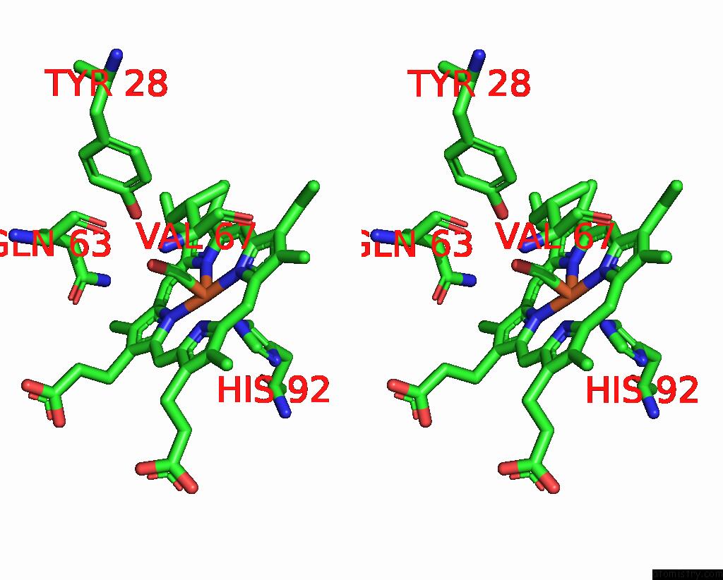
Iron binding site 2 out of 4 in 1j7y
Go back to
Iron binding site 2 out
of 4 in the Crystal Structure of Partially Ligated Mutant of Hba
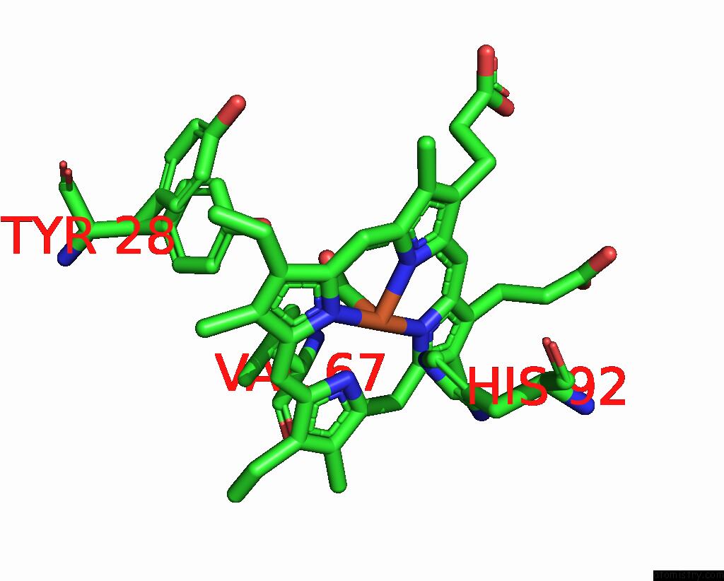
Mono view
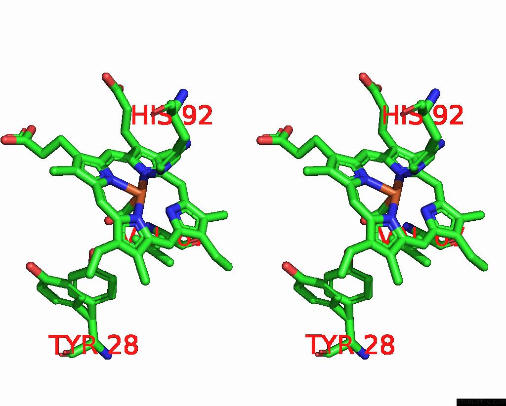
Stereo pair view
Mono view
Stereo pair view
A full contact list of Iron with other atoms in the Fe binding
site number 2 of Crystal Structure of Partially Ligated Mutant of Hba within 5.0Å range:
|
Iron binding site 3 out of 4 in 1j7y
Go back to
Iron binding site 3 out
of 4 in the Crystal Structure of Partially Ligated Mutant of Hba
Mono view
Stereo pair view
Mono view
Stereo pair view
A full contact list of Iron with other atoms in the Fe binding
site number 3 of Crystal Structure of Partially Ligated Mutant of Hba within 5.0Å range:
|
Iron binding site 4 out of 4 in 1j7y
Go back to
Iron binding site 4 out
of 4 in the Crystal Structure of Partially Ligated Mutant of Hba
Mono view
Stereo pair view
Mono view
Stereo pair view
A full contact list of Iron with other atoms in the Fe binding
site number 4 of Crystal Structure of Partially Ligated Mutant of Hba within 5.0Å range:
|
Reference:
A.E.Miele,
F.Draghi,
A.Arcovito,
A.Bellelli,
M.Brunori,
C.Travaglini-Allocatelli,
B.Vallone.
Control of Heme Reactivity By Diffusion: Structural Basis and Functional Characterization in Hemoglobin Mutants. Biochemistry V. 40 14449 2001.
ISSN: ISSN 0006-2960
PubMed: 11724557
DOI: 10.1021/BI011602D
Page generated: Wed Jul 16 16:28:46 2025
ISSN: ISSN 0006-2960
PubMed: 11724557
DOI: 10.1021/BI011602D
Last articles
Fe in 1U9UFe in 1UB2
Fe in 1U7R
Fe in 1U7S
Fe in 1U74
Fe in 1U5U
Fe in 1U75
Fe in 1U56
Fe in 1U55
Fe in 1U4H