Iron »
PDB 1kqj-1lfg »
1kqj »
Iron in PDB 1kqj: Crystal Structure of A Mutant of Muty Catalytic Domain
Protein crystallography data
The structure of Crystal Structure of A Mutant of Muty Catalytic Domain, PDB code: 1kqj
was solved by
T.E.Messick,
N.H.Chmiel,
M.P.Golinelli,
S.S.David,
L.Joshua-Tor,
with X-Ray Crystallography technique. A brief refinement statistics is given in the table below:
| Resolution Low / High (Å) | 29.65 / 1.70 |
| Space group | C 1 2 1 |
| Cell size a, b, c (Å), α, β, γ (°) | 83.550, 49.900, 71.010, 90.00, 122.56, 90.00 |
| R / Rfree (%) | 19.1 / 20.8 |
Iron Binding Sites:
The binding sites of Iron atom in the Crystal Structure of A Mutant of Muty Catalytic Domain
(pdb code 1kqj). This binding sites where shown within
5.0 Angstroms radius around Iron atom.
In total 4 binding sites of Iron where determined in the Crystal Structure of A Mutant of Muty Catalytic Domain, PDB code: 1kqj:
Jump to Iron binding site number: 1; 2; 3; 4;
In total 4 binding sites of Iron where determined in the Crystal Structure of A Mutant of Muty Catalytic Domain, PDB code: 1kqj:
Jump to Iron binding site number: 1; 2; 3; 4;
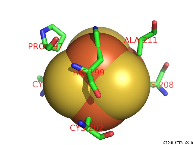
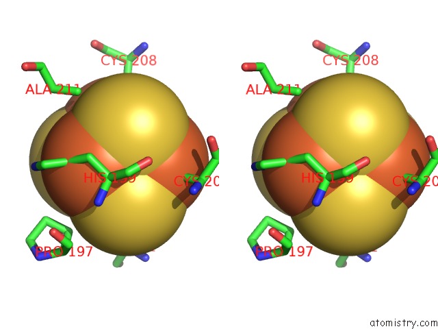
Iron binding site 1 out of 4 in 1kqj
Go back to
Iron binding site 1 out
of 4 in the Crystal Structure of A Mutant of Muty Catalytic Domain
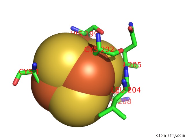
Mono view
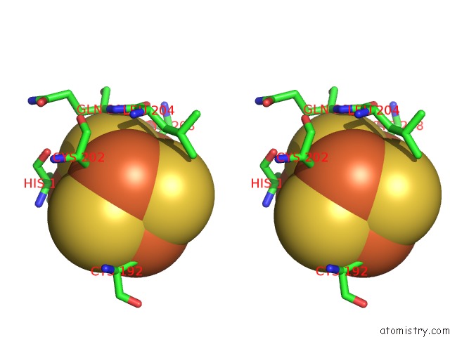
Stereo pair view
Mono view
Stereo pair view
A full contact list of Iron with other atoms in the Fe binding
site number 1 of Crystal Structure of A Mutant of Muty Catalytic Domain within 5.0Å range:
|
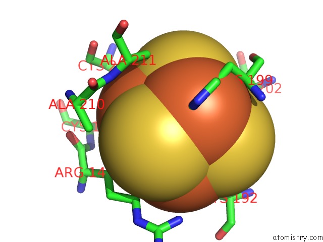
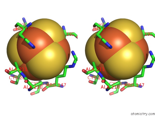
Iron binding site 2 out of 4 in 1kqj
Go back to
Iron binding site 2 out
of 4 in the Crystal Structure of A Mutant of Muty Catalytic Domain
Mono view
Stereo pair view
Mono view
Stereo pair view
A full contact list of Iron with other atoms in the Fe binding
site number 2 of Crystal Structure of A Mutant of Muty Catalytic Domain within 5.0Å range:
|
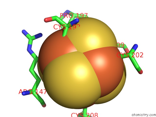
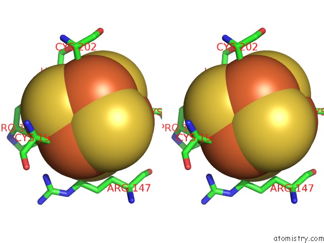
Iron binding site 3 out of 4 in 1kqj
Go back to
Iron binding site 3 out
of 4 in the Crystal Structure of A Mutant of Muty Catalytic Domain
Mono view
Stereo pair view
Mono view
Stereo pair view
A full contact list of Iron with other atoms in the Fe binding
site number 3 of Crystal Structure of A Mutant of Muty Catalytic Domain within 5.0Å range:
|
Iron binding site 4 out of 4 in 1kqj
Go back to
Iron binding site 4 out
of 4 in the Crystal Structure of A Mutant of Muty Catalytic Domain
Mono view
Stereo pair view
Mono view
Stereo pair view
A full contact list of Iron with other atoms in the Fe binding
site number 4 of Crystal Structure of A Mutant of Muty Catalytic Domain within 5.0Å range:
|
Reference:
T.E.Messick,
N.H.Chmiel,
M.P.Golinelli,
M.R.Langer,
L.Joshua-Tor,
S.S.David.
Noncysteinyl Coordination to the [4FE-4S]2+ Cluster of the Dna Repair Adenine Glycosylase Muty Introduced Via Site-Directed Mutagenesis. Structural Characterization of An Unusual Histidinyl-Coordinated Cluster. Biochemistry V. 41 3931 2002.
ISSN: ISSN 0006-2960
PubMed: 11900536
DOI: 10.1021/BI012035X
Page generated: Wed Jul 16 17:11:58 2025
ISSN: ISSN 0006-2960
PubMed: 11900536
DOI: 10.1021/BI012035X
Last articles
Fe in 2YXOFe in 2YRS
Fe in 2YXC
Fe in 2YNM
Fe in 2YVJ
Fe in 2YP1
Fe in 2YU2
Fe in 2YU1
Fe in 2YQB
Fe in 2YOO