Iron »
PDB 1kqj-1lfg »
1kr7 »
Iron in PDB 1kr7: Crystal Structure of the Nerve Tissue Mini-Hemoglobin From the Nemertean Worm Cerebratulus Lacteus
Protein crystallography data
The structure of Crystal Structure of the Nerve Tissue Mini-Hemoglobin From the Nemertean Worm Cerebratulus Lacteus, PDB code: 1kr7
was solved by
A.Pesce,
M.Nardini,
S.Dewilde,
E.Geuens,
K.Yamauchi,
P.Ascenzi,
A.F.Riggs,
L.Moens,
M.Bolognesi,
with X-Ray Crystallography technique. A brief refinement statistics is given in the table below:
| Resolution Low / High (Å) | 35.14 / 1.50 |
| Space group | P 21 21 21 |
| Cell size a, b, c (Å), α, β, γ (°) | 42.721, 43.173, 60.110, 90.00, 90.00, 90.00 |
| R / Rfree (%) | 15.3 / 18.7 |
Iron Binding Sites:
The binding sites of Iron atom in the Crystal Structure of the Nerve Tissue Mini-Hemoglobin From the Nemertean Worm Cerebratulus Lacteus
(pdb code 1kr7). This binding sites where shown within
5.0 Angstroms radius around Iron atom.
In total only one binding site of Iron was determined in the Crystal Structure of the Nerve Tissue Mini-Hemoglobin From the Nemertean Worm Cerebratulus Lacteus, PDB code: 1kr7:
In total only one binding site of Iron was determined in the Crystal Structure of the Nerve Tissue Mini-Hemoglobin From the Nemertean Worm Cerebratulus Lacteus, PDB code: 1kr7:
Iron binding site 1 out of 1 in 1kr7
Go back to
Iron binding site 1 out
of 1 in the Crystal Structure of the Nerve Tissue Mini-Hemoglobin From the Nemertean Worm Cerebratulus Lacteus
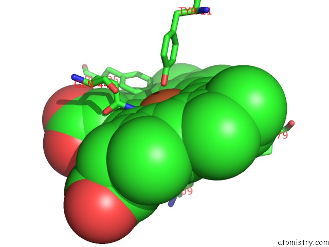
Mono view
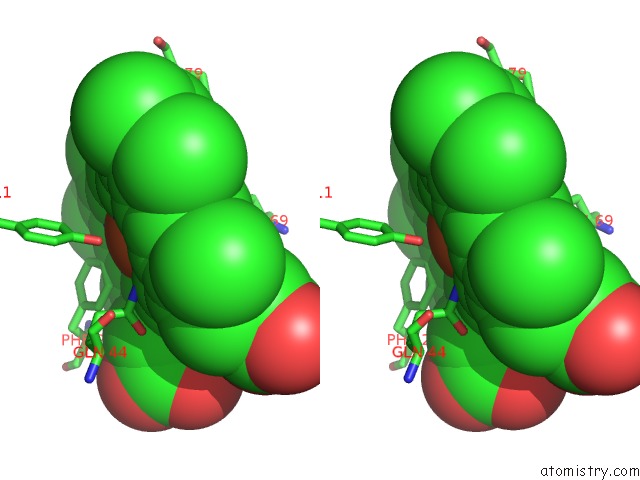
Stereo pair view
Mono view
Stereo pair view
A full contact list of Iron with other atoms in the Fe binding
site number 1 of Crystal Structure of the Nerve Tissue Mini-Hemoglobin From the Nemertean Worm Cerebratulus Lacteus within 5.0Å range:
|
Reference:
A.Pesce,
M.Nardini,
S.Dewilde,
E.Geuens,
K.Yamauchi,
P.Ascenzi,
A.F.Riggs,
L.Moens,
M.Bolognesi.
The 109 Residue Nerve Tissue Minihemoglobin From Cerebratulus Lacteus Highlights Striking Structural Plasticity of the Alpha-Helical Globin Fold Structure V. 10 725 2002.
ISSN: ISSN 0969-2126
PubMed: 12015154
DOI: 10.1016/S0969-2126(02)00763-3
Page generated: Sat Aug 3 09:29:22 2024
ISSN: ISSN 0969-2126
PubMed: 12015154
DOI: 10.1016/S0969-2126(02)00763-3
Last articles
Zn in 9MJ5Zn in 9HNW
Zn in 9G0L
Zn in 9FNE
Zn in 9DZN
Zn in 9E0I
Zn in 9D32
Zn in 9DAK
Zn in 8ZXC
Zn in 8ZUF