Iron »
PDB 1q5e-1qom »
1qhu »
Iron in PDB 1qhu: Mammalian Blood Serum Haemopexin Deglycosylated and in Complex with Its Ligand Haem
Protein crystallography data
The structure of Mammalian Blood Serum Haemopexin Deglycosylated and in Complex with Its Ligand Haem, PDB code: 1qhu
was solved by
M.Paoli,
H.M.Baker,
W.T.Morgan,
A.Smith,
E.N.Baker,
with X-Ray Crystallography technique. A brief refinement statistics is given in the table below:
| Resolution Low / High (Å) | 20.00 / 2.30 |
| Space group | P 1 21 1 |
| Cell size a, b, c (Å), α, β, γ (°) | 44.680, 61.950, 83.290, 90.00, 93.21, 90.00 |
| R / Rfree (%) | 20.7 / 28.9 |
Other elements in 1qhu:
The structure of Mammalian Blood Serum Haemopexin Deglycosylated and in Complex with Its Ligand Haem also contains other interesting chemical elements:
| Chlorine | (Cl) | 2 atoms |
| Sodium | (Na) | 4 atoms |
Iron Binding Sites:
The binding sites of Iron atom in the Mammalian Blood Serum Haemopexin Deglycosylated and in Complex with Its Ligand Haem
(pdb code 1qhu). This binding sites where shown within
5.0 Angstroms radius around Iron atom.
In total only one binding site of Iron was determined in the Mammalian Blood Serum Haemopexin Deglycosylated and in Complex with Its Ligand Haem, PDB code: 1qhu:
In total only one binding site of Iron was determined in the Mammalian Blood Serum Haemopexin Deglycosylated and in Complex with Its Ligand Haem, PDB code: 1qhu:
Iron binding site 1 out of 1 in 1qhu
Go back to
Iron binding site 1 out
of 1 in the Mammalian Blood Serum Haemopexin Deglycosylated and in Complex with Its Ligand Haem
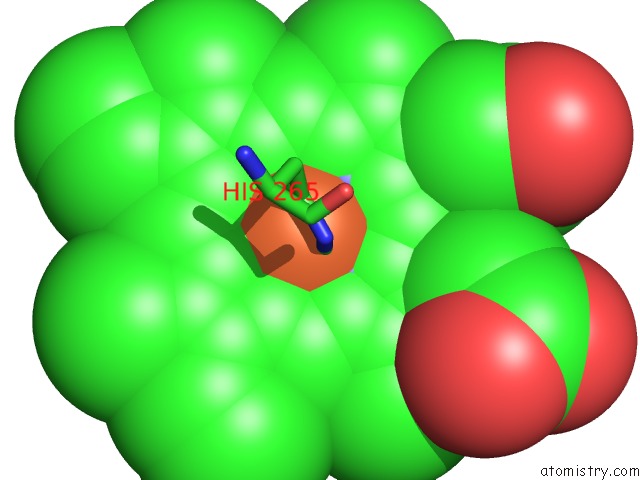
Mono view
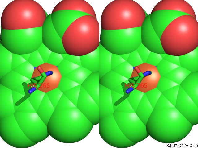
Stereo pair view
Mono view
Stereo pair view
A full contact list of Iron with other atoms in the Fe binding
site number 1 of Mammalian Blood Serum Haemopexin Deglycosylated and in Complex with Its Ligand Haem within 5.0Å range:
|
Reference:
M.Paoli,
B.F.Anderson,
H.M.Baker,
W.T.Morgan,
A.Smith,
E.N.Baker.
Crystal Structure of Hemopexin Reveals A Novel High-Affinity Heme Site Formed Between Two Beta-Propeller Domains. Nat.Struct.Biol. V. 6 926 1999.
ISSN: ISSN 1072-8368
PubMed: 10504726
DOI: 10.1038/13294
Page generated: Sat Aug 3 13:26:37 2024
ISSN: ISSN 1072-8368
PubMed: 10504726
DOI: 10.1038/13294
Last articles
Zn in 9J0NZn in 9J0O
Zn in 9J0P
Zn in 9FJX
Zn in 9EKB
Zn in 9C0F
Zn in 9CAH
Zn in 9CH0
Zn in 9CH3
Zn in 9CH1