Iron »
PDB 1q5e-1qom »
1qn2 »
Iron in PDB 1qn2: Cytochrome Ch From Methylobacterium Extorquens
Protein crystallography data
The structure of Cytochrome Ch From Methylobacterium Extorquens, PDB code: 1qn2
was solved by
J.Read,
R.Gill,
S.L.Dales,
J.B.Cooper,
S.P.Wood,
C.Anthony,
with X-Ray Crystallography technique. A brief refinement statistics is given in the table below:
| Resolution Low / High (Å) | 30.00 / 2.01 |
| Space group | P 1 |
| Cell size a, b, c (Å), α, β, γ (°) | 33.760, 57.570, 50.950, 67.81, 89.33, 74.40 |
| R / Rfree (%) | n/a / n/a |
Iron Binding Sites:
The binding sites of Iron atom in the Cytochrome Ch From Methylobacterium Extorquens
(pdb code 1qn2). This binding sites where shown within
5.0 Angstroms radius around Iron atom.
In total 3 binding sites of Iron where determined in the Cytochrome Ch From Methylobacterium Extorquens, PDB code: 1qn2:
Jump to Iron binding site number: 1; 2; 3;
In total 3 binding sites of Iron where determined in the Cytochrome Ch From Methylobacterium Extorquens, PDB code: 1qn2:
Jump to Iron binding site number: 1; 2; 3;
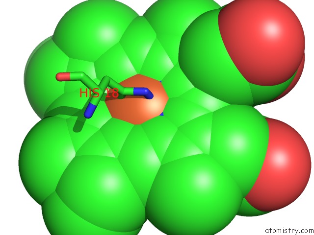
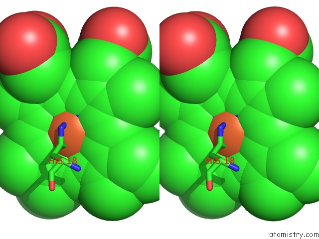
Iron binding site 1 out of 3 in 1qn2
Go back to
Iron binding site 1 out
of 3 in the Cytochrome Ch From Methylobacterium Extorquens
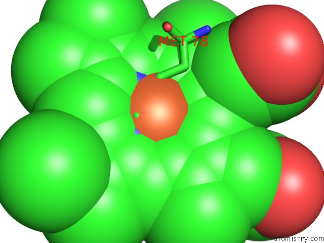
Mono view
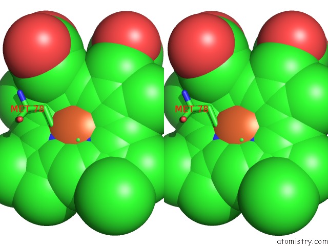
Stereo pair view
Mono view
Stereo pair view
A full contact list of Iron with other atoms in the Fe binding
site number 1 of Cytochrome Ch From Methylobacterium Extorquens within 5.0Å range:
|
Iron binding site 2 out of 3 in 1qn2
Go back to
Iron binding site 2 out
of 3 in the Cytochrome Ch From Methylobacterium Extorquens
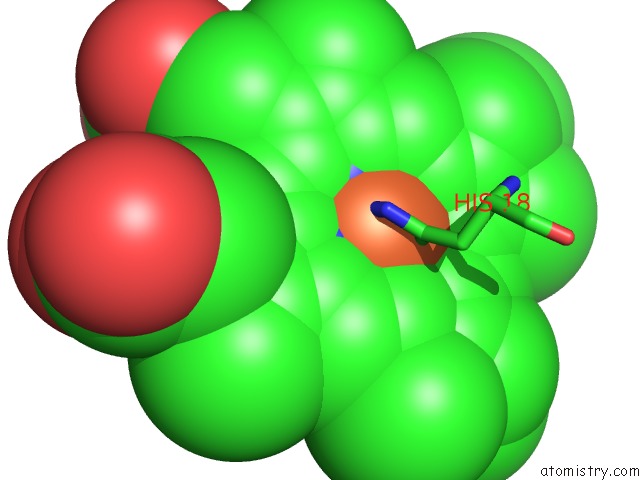
Mono view
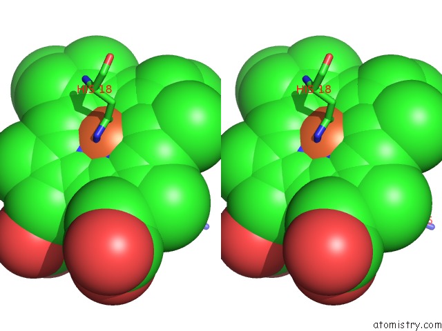
Stereo pair view
Mono view
Stereo pair view
A full contact list of Iron with other atoms in the Fe binding
site number 2 of Cytochrome Ch From Methylobacterium Extorquens within 5.0Å range:
|
Iron binding site 3 out of 3 in 1qn2
Go back to
Iron binding site 3 out
of 3 in the Cytochrome Ch From Methylobacterium Extorquens
Mono view
Stereo pair view
Mono view
Stereo pair view
A full contact list of Iron with other atoms in the Fe binding
site number 3 of Cytochrome Ch From Methylobacterium Extorquens within 5.0Å range:
|
Reference:
J.Read,
R.Gill,
S.L.Dales,
J.B.Cooper,
S.P.Wood,
C.Anthony.
The Molecular Structure of An Unusual Cytochrome C2 Determined at 2.0A; the Cytochrome Ch From Methylobacterium Extorquens Protein Sci. V. 8 1232 1999.
ISSN: ISSN 0961-8368
PubMed: 10386873
DOI: 10.1110/PS.8.6.1232
Page generated: Wed Jul 16 20:02:59 2025
ISSN: ISSN 0961-8368
PubMed: 10386873
DOI: 10.1110/PS.8.6.1232
Last articles
Fe in 2YXOFe in 2YRS
Fe in 2YXC
Fe in 2YNM
Fe in 2YVJ
Fe in 2YP1
Fe in 2YU2
Fe in 2YU1
Fe in 2YQB
Fe in 2YOO