Iron »
PDB 1q5e-1qom »
1qof »
Iron in PDB 1qof: Ferredoxin Mutation Q70K
Protein crystallography data
The structure of Ferredoxin Mutation Q70K, PDB code: 1qof
was solved by
H.M.Holden,
M.M.Benning,
with X-Ray Crystallography technique. A brief refinement statistics is given in the table below:
| Resolution Low / High (Å) | 25.00 / 1.80 |
| Space group | P 21 21 21 |
| Cell size a, b, c (Å), α, β, γ (°) | 37.730, 38.230, 148.160, 90.00, 90.00, 90.00 |
| R / Rfree (%) | 17.4 / n/a |
Iron Binding Sites:
The binding sites of Iron atom in the Ferredoxin Mutation Q70K
(pdb code 1qof). This binding sites where shown within
5.0 Angstroms radius around Iron atom.
In total 4 binding sites of Iron where determined in the Ferredoxin Mutation Q70K, PDB code: 1qof:
Jump to Iron binding site number: 1; 2; 3; 4;
In total 4 binding sites of Iron where determined in the Ferredoxin Mutation Q70K, PDB code: 1qof:
Jump to Iron binding site number: 1; 2; 3; 4;
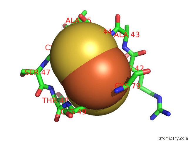
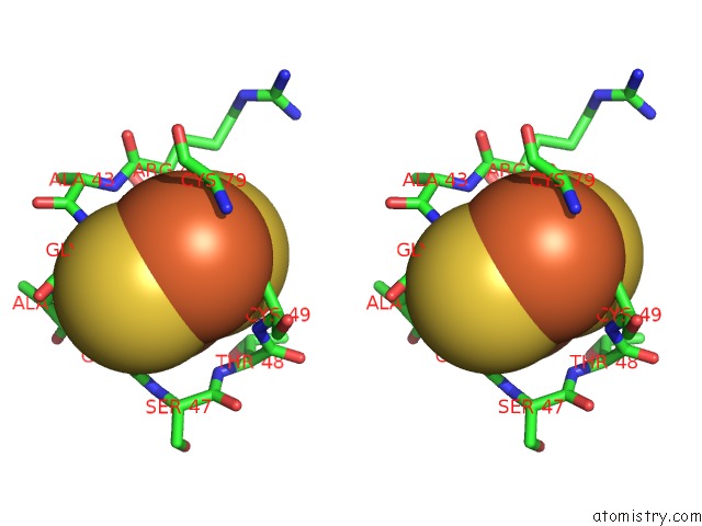
Iron binding site 1 out of 4 in 1qof
Go back to
Iron binding site 1 out
of 4 in the Ferredoxin Mutation Q70K
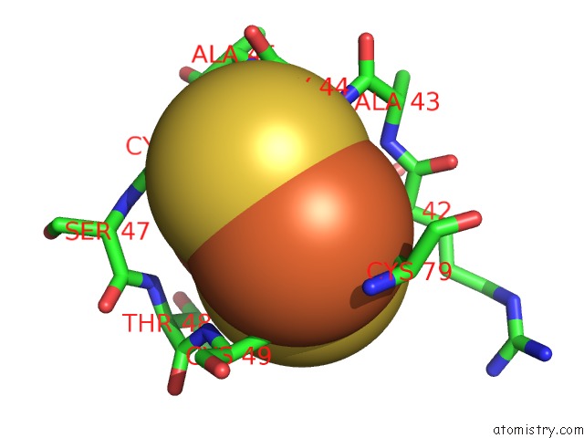
Mono view
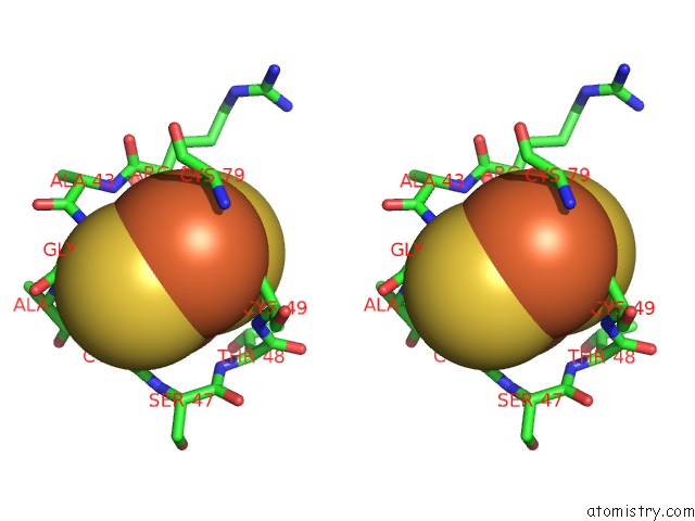
Stereo pair view
Mono view
Stereo pair view
A full contact list of Iron with other atoms in the Fe binding
site number 1 of Ferredoxin Mutation Q70K within 5.0Å range:
|
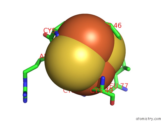
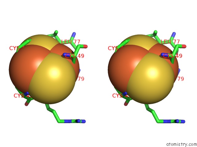
Iron binding site 2 out of 4 in 1qof
Go back to
Iron binding site 2 out
of 4 in the Ferredoxin Mutation Q70K
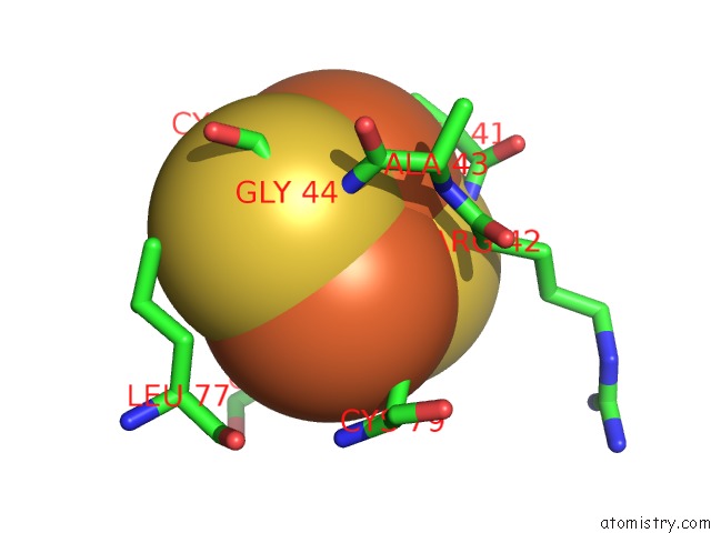
Mono view
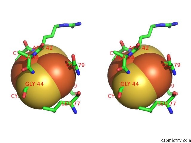
Stereo pair view
Mono view
Stereo pair view
A full contact list of Iron with other atoms in the Fe binding
site number 2 of Ferredoxin Mutation Q70K within 5.0Å range:
|
Iron binding site 3 out of 4 in 1qof
Go back to
Iron binding site 3 out
of 4 in the Ferredoxin Mutation Q70K
Mono view
Stereo pair view
Mono view
Stereo pair view
A full contact list of Iron with other atoms in the Fe binding
site number 3 of Ferredoxin Mutation Q70K within 5.0Å range:
|
Iron binding site 4 out of 4 in 1qof
Go back to
Iron binding site 4 out
of 4 in the Ferredoxin Mutation Q70K
Mono view
Stereo pair view
Mono view
Stereo pair view
A full contact list of Iron with other atoms in the Fe binding
site number 4 of Ferredoxin Mutation Q70K within 5.0Å range:
|
Reference:
J.K.Hurley,
A.M.Weber-Main,
M.T.Stankovich,
M.M.Benning,
J.B.Thoden,
J.L.Vanhooke,
H.M.Holden,
Y.K.Chae,
B.Xia,
H.Cheng,
J.L.Markley,
M.Martinez-Julvez,
C.Gomez-Moreno,
J.L.Schmeits,
G.Tollin.
Structure-Function Relationships in Anabaena Ferredoxin: Correlations Between X-Ray Crystal Structures, Reduction Potentials, and Rate Constants of Electron Transfer to Ferredoxin:Nadp+ Reductase For Site-Specific Ferredoxin Mutants. Biochemistry V. 36 11100 1997.
ISSN: ISSN 0006-2960
PubMed: 9287153
DOI: 10.1021/BI9709001
Page generated: Sat Aug 3 13:40:19 2024
ISSN: ISSN 0006-2960
PubMed: 9287153
DOI: 10.1021/BI9709001
Last articles
Zn in 9MJ5Zn in 9HNW
Zn in 9G0L
Zn in 9FNE
Zn in 9DZN
Zn in 9E0I
Zn in 9D32
Zn in 9DAK
Zn in 8ZXC
Zn in 8ZUF