Iron »
PDB 1sdl-1stq »
1sdq »
Iron in PDB 1sdq: Structure of Reduced-No Adduct of Mesopone Cytochrome C Peroxidase
Enzymatic activity of Structure of Reduced-No Adduct of Mesopone Cytochrome C Peroxidase
All present enzymatic activity of Structure of Reduced-No Adduct of Mesopone Cytochrome C Peroxidase:
1.11.1.5;
1.11.1.5;
Protein crystallography data
The structure of Structure of Reduced-No Adduct of Mesopone Cytochrome C Peroxidase, PDB code: 1sdq
was solved by
B.Bhaskar,
C.E.Immoos,
F.Sulc,
M.S.Cohem,
P.J.Farmer,
T.L.Poulos,
with X-Ray Crystallography technique. A brief refinement statistics is given in the table below:
| Resolution Low / High (Å) | 50.00 / 1.69 |
| Space group | P 21 21 21 |
| Cell size a, b, c (Å), α, β, γ (°) | 106.884, 75.625, 50.958, 90.00, 90.00, 90.00 |
| R / Rfree (%) | 19.3 / 21.5 |
Iron Binding Sites:
The binding sites of Iron atom in the Structure of Reduced-No Adduct of Mesopone Cytochrome C Peroxidase
(pdb code 1sdq). This binding sites where shown within
5.0 Angstroms radius around Iron atom.
In total only one binding site of Iron was determined in the Structure of Reduced-No Adduct of Mesopone Cytochrome C Peroxidase, PDB code: 1sdq:
In total only one binding site of Iron was determined in the Structure of Reduced-No Adduct of Mesopone Cytochrome C Peroxidase, PDB code: 1sdq:
Iron binding site 1 out of 1 in 1sdq
Go back to
Iron binding site 1 out
of 1 in the Structure of Reduced-No Adduct of Mesopone Cytochrome C Peroxidase
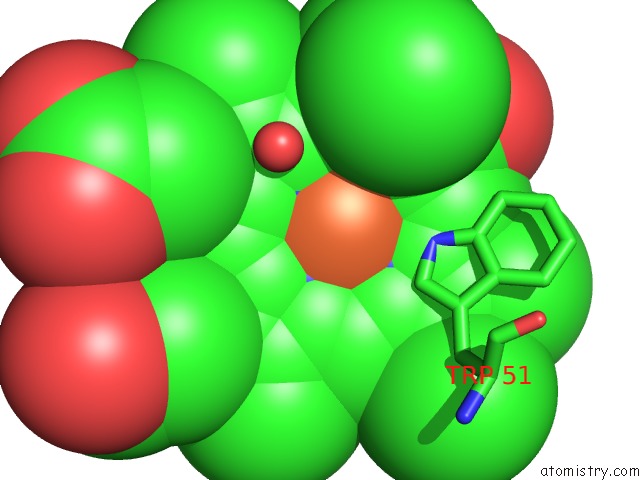
Mono view
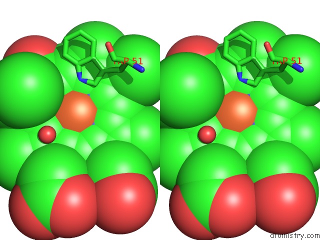
Stereo pair view
Mono view
Stereo pair view
A full contact list of Iron with other atoms in the Fe binding
site number 1 of Structure of Reduced-No Adduct of Mesopone Cytochrome C Peroxidase within 5.0Å range:
|
Reference:
B.Bhaskar,
C.E.Immoos,
F.Sulc,
M.S.Cohem,
P.J.Farmer,
T.L.Poulos.
Crystal Structures of Resting (FE3+), Reduced (FE2+) and No-Bound States of Mesopone Cytochrome C Peroxidase (Mpccp) (R-Isomer) To Be Published.
Page generated: Wed Jul 16 20:37:56 2025
Last articles
Fe in 2YXOFe in 2YRS
Fe in 2YXC
Fe in 2YNM
Fe in 2YVJ
Fe in 2YP1
Fe in 2YU2
Fe in 2YU1
Fe in 2YQB
Fe in 2YOO