Iron »
PDB 1sdl-1stq »
1se6 »
Iron in PDB 1se6: Crystal Structure of Streptomyces Coelicolor A3(2) CYP158A2 From Antibiotic Biosynthetic Pathways
Protein crystallography data
The structure of Crystal Structure of Streptomyces Coelicolor A3(2) CYP158A2 From Antibiotic Biosynthetic Pathways, PDB code: 1se6
was solved by
B.Zhao,
D.C.Lamb,
L.Lei,
M.Sundaramoorthy,
L.M.Podust,
M.R.Waterman,
with X-Ray Crystallography technique. A brief refinement statistics is given in the table below:
| Resolution Low / High (Å) | 35.90 / 1.75 |
| Space group | P 1 21 1 |
| Cell size a, b, c (Å), α, β, γ (°) | 59.554, 79.206, 87.430, 90.00, 92.26, 90.00 |
| R / Rfree (%) | 23.4 / 26.8 |
Iron Binding Sites:
The binding sites of Iron atom in the Crystal Structure of Streptomyces Coelicolor A3(2) CYP158A2 From Antibiotic Biosynthetic Pathways
(pdb code 1se6). This binding sites where shown within
5.0 Angstroms radius around Iron atom.
In total 2 binding sites of Iron where determined in the Crystal Structure of Streptomyces Coelicolor A3(2) CYP158A2 From Antibiotic Biosynthetic Pathways, PDB code: 1se6:
Jump to Iron binding site number: 1; 2;
In total 2 binding sites of Iron where determined in the Crystal Structure of Streptomyces Coelicolor A3(2) CYP158A2 From Antibiotic Biosynthetic Pathways, PDB code: 1se6:
Jump to Iron binding site number: 1; 2;
Iron binding site 1 out of 2 in 1se6
Go back to
Iron binding site 1 out
of 2 in the Crystal Structure of Streptomyces Coelicolor A3(2) CYP158A2 From Antibiotic Biosynthetic Pathways
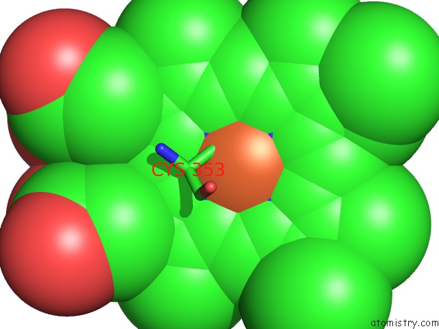
Mono view
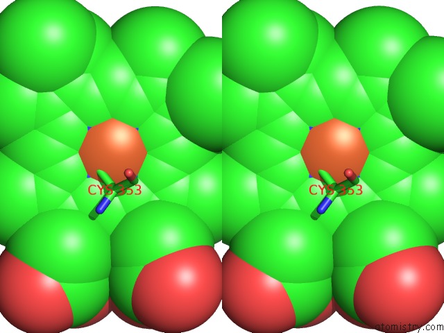
Stereo pair view
Mono view
Stereo pair view
A full contact list of Iron with other atoms in the Fe binding
site number 1 of Crystal Structure of Streptomyces Coelicolor A3(2) CYP158A2 From Antibiotic Biosynthetic Pathways within 5.0Å range:
|
Iron binding site 2 out of 2 in 1se6
Go back to
Iron binding site 2 out
of 2 in the Crystal Structure of Streptomyces Coelicolor A3(2) CYP158A2 From Antibiotic Biosynthetic Pathways
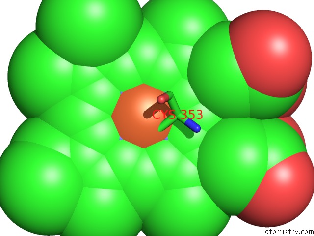
Mono view
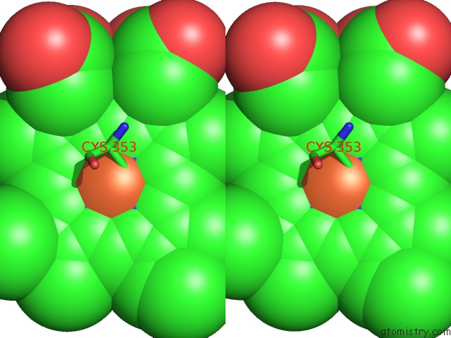
Stereo pair view
Mono view
Stereo pair view
A full contact list of Iron with other atoms in the Fe binding
site number 2 of Crystal Structure of Streptomyces Coelicolor A3(2) CYP158A2 From Antibiotic Biosynthetic Pathways within 5.0Å range:
|
Reference:
B.Zhao,
F.P.Guengerich,
A.Bellamine,
D.C.Lamb,
M.Izumikawa,
L.Lei,
L.M.Podust,
M.Sundaramoorthy,
J.A.Kalaitzis,
L.M.Reddy,
S.L.Kelly,
B.S.Moore,
D.Stec,
M.Voehler,
J.R.Falck,
T.Shimada,
M.R.Waterman.
Binding of Two Flaviolin Substrate Molecules, Oxidative Coupling, and Crystal Structure of Streptomyces Coelicolor A3(2) Cytochrome P450 158A2. J.Biol.Chem. V. 280 11599 2005.
ISSN: ISSN 0021-9258
PubMed: 15659395
DOI: 10.1074/JBC.M410933200
Page generated: Wed Jul 16 20:37:56 2025
ISSN: ISSN 0021-9258
PubMed: 15659395
DOI: 10.1074/JBC.M410933200
Last articles
Fe in 2YXOFe in 2YRS
Fe in 2YXC
Fe in 2YNM
Fe in 2YVJ
Fe in 2YP1
Fe in 2YU2
Fe in 2YU1
Fe in 2YQB
Fe in 2YOO