Iron »
PDB 1sdl-1stq »
1shr »
Iron in PDB 1shr: Crystal Structure of Ferrocyanide Bound Human Hemoglobin A2 at 1.88A Resolution
Protein crystallography data
The structure of Crystal Structure of Ferrocyanide Bound Human Hemoglobin A2 at 1.88A Resolution, PDB code: 1shr
was solved by
U.Sen,
J.Dasgupta,
D.Choudhury,
P.Datta,
A.Chakrabarti,
S.B.Chakrabarty,
A.Chakrabarty,
J.K.Dattagupta,
with X-Ray Crystallography technique. A brief refinement statistics is given in the table below:
| Resolution Low / High (Å) | 14.89 / 1.88 |
| Space group | P 1 21 1 |
| Cell size a, b, c (Å), α, β, γ (°) | 54.449, 83.990, 62.688, 90.00, 99.86, 90.00 |
| R / Rfree (%) | 16.7 / 19.5 |
Iron Binding Sites:
The binding sites of Iron atom in the Crystal Structure of Ferrocyanide Bound Human Hemoglobin A2 at 1.88A Resolution
(pdb code 1shr). This binding sites where shown within
5.0 Angstroms radius around Iron atom.
In total 7 binding sites of Iron where determined in the Crystal Structure of Ferrocyanide Bound Human Hemoglobin A2 at 1.88A Resolution, PDB code: 1shr:
Jump to Iron binding site number: 1; 2; 3; 4; 5; 6; 7;
In total 7 binding sites of Iron where determined in the Crystal Structure of Ferrocyanide Bound Human Hemoglobin A2 at 1.88A Resolution, PDB code: 1shr:
Jump to Iron binding site number: 1; 2; 3; 4; 5; 6; 7;
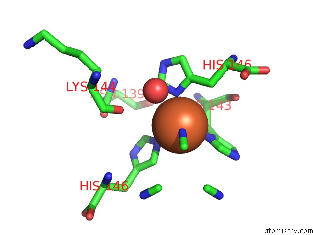
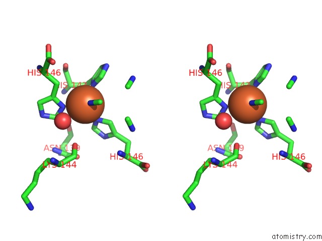
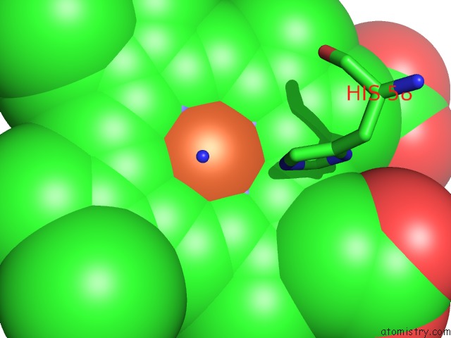
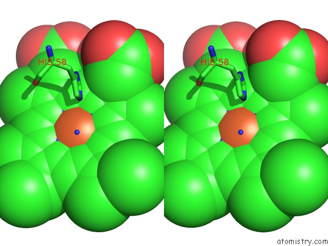
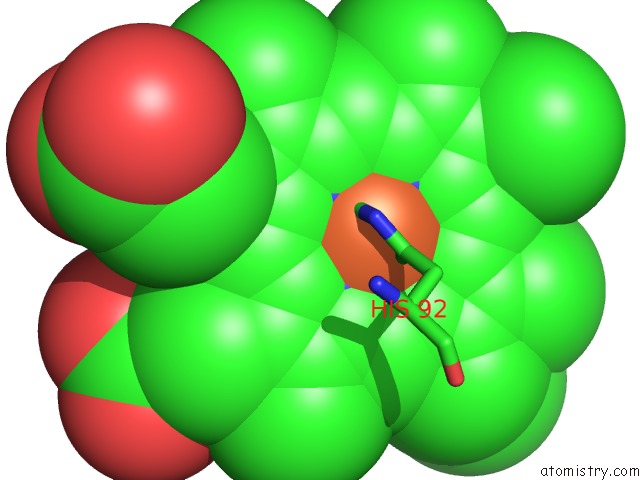
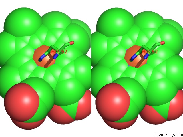
Iron binding site 1 out of 7 in 1shr
Go back to
Iron binding site 1 out
of 7 in the Crystal Structure of Ferrocyanide Bound Human Hemoglobin A2 at 1.88A Resolution
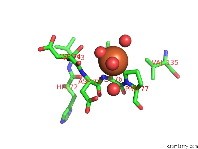
Mono view
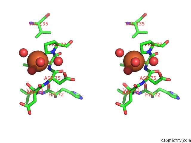
Stereo pair view
Mono view
Stereo pair view
A full contact list of Iron with other atoms in the Fe binding
site number 1 of Crystal Structure of Ferrocyanide Bound Human Hemoglobin A2 at 1.88A Resolution within 5.0Å range:
|
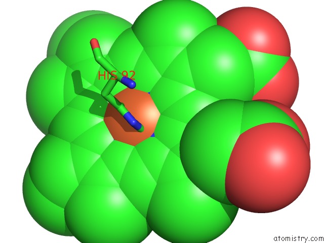
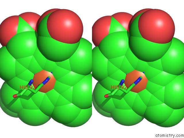
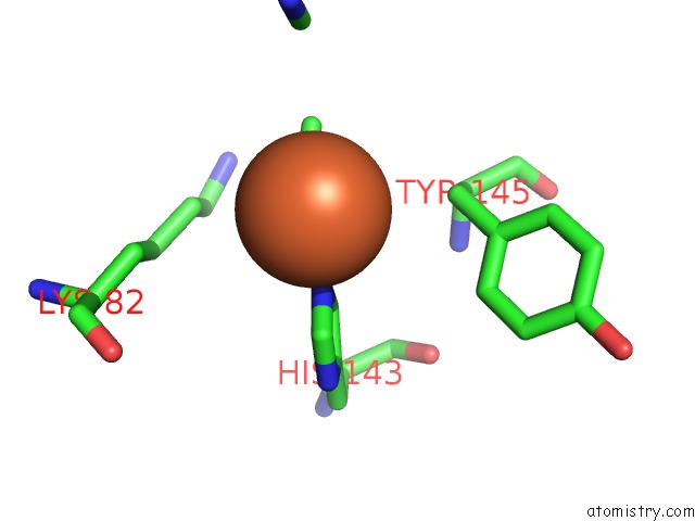
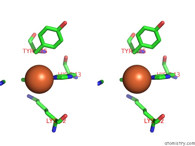
Iron binding site 2 out of 7 in 1shr
Go back to
Iron binding site 2 out
of 7 in the Crystal Structure of Ferrocyanide Bound Human Hemoglobin A2 at 1.88A Resolution
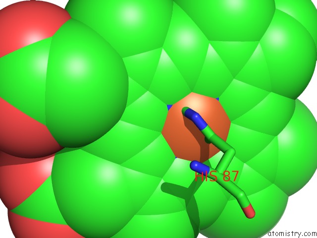
Mono view
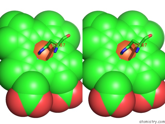
Stereo pair view
Mono view
Stereo pair view
A full contact list of Iron with other atoms in the Fe binding
site number 2 of Crystal Structure of Ferrocyanide Bound Human Hemoglobin A2 at 1.88A Resolution within 5.0Å range:
|
Iron binding site 3 out of 7 in 1shr
Go back to
Iron binding site 3 out
of 7 in the Crystal Structure of Ferrocyanide Bound Human Hemoglobin A2 at 1.88A Resolution
Mono view
Stereo pair view
Mono view
Stereo pair view
A full contact list of Iron with other atoms in the Fe binding
site number 3 of Crystal Structure of Ferrocyanide Bound Human Hemoglobin A2 at 1.88A Resolution within 5.0Å range:
|
Iron binding site 4 out of 7 in 1shr
Go back to
Iron binding site 4 out
of 7 in the Crystal Structure of Ferrocyanide Bound Human Hemoglobin A2 at 1.88A Resolution
Mono view
Stereo pair view
Mono view
Stereo pair view
A full contact list of Iron with other atoms in the Fe binding
site number 4 of Crystal Structure of Ferrocyanide Bound Human Hemoglobin A2 at 1.88A Resolution within 5.0Å range:
|
Iron binding site 5 out of 7 in 1shr
Go back to
Iron binding site 5 out
of 7 in the Crystal Structure of Ferrocyanide Bound Human Hemoglobin A2 at 1.88A Resolution
Mono view
Stereo pair view
Mono view
Stereo pair view
A full contact list of Iron with other atoms in the Fe binding
site number 5 of Crystal Structure of Ferrocyanide Bound Human Hemoglobin A2 at 1.88A Resolution within 5.0Å range:
|
Iron binding site 6 out of 7 in 1shr
Go back to
Iron binding site 6 out
of 7 in the Crystal Structure of Ferrocyanide Bound Human Hemoglobin A2 at 1.88A Resolution
Mono view
Stereo pair view
Mono view
Stereo pair view
A full contact list of Iron with other atoms in the Fe binding
site number 6 of Crystal Structure of Ferrocyanide Bound Human Hemoglobin A2 at 1.88A Resolution within 5.0Å range:
|
Iron binding site 7 out of 7 in 1shr
Go back to
Iron binding site 7 out
of 7 in the Crystal Structure of Ferrocyanide Bound Human Hemoglobin A2 at 1.88A Resolution
Mono view
Stereo pair view
Mono view
Stereo pair view
A full contact list of Iron with other atoms in the Fe binding
site number 7 of Crystal Structure of Ferrocyanide Bound Human Hemoglobin A2 at 1.88A Resolution within 5.0Å range:
|
Reference:
U.Sen,
J.Dasgupta,
D.Choudhury,
P.Datta,
A.Chakrabarti,
S.B.Chakrabarty,
A.Chakrabarty,
J.K.Dattagupta.
Crystal Structures of HBA2 and Hbe and Modeling of Hemoglobin DELTA4: Interpretation of the Thermal Stability and the Antisickling Effect of HBA2 and Identification of the Ferrocyanide Binding Site in Hb Biochemistry V. 43 12477 2004.
ISSN: ISSN 0006-2960
PubMed: 15449937
DOI: 10.1021/BI048903I
Page generated: Sat Aug 3 14:41:19 2024
ISSN: ISSN 0006-2960
PubMed: 15449937
DOI: 10.1021/BI048903I
Last articles
Zn in 9MJ5Zn in 9HNW
Zn in 9G0L
Zn in 9FNE
Zn in 9DZN
Zn in 9E0I
Zn in 9D32
Zn in 9DAK
Zn in 8ZXC
Zn in 8ZUF